Zhang Qiaoli, Ding Yingxue, Yao Yanqing, Yu Yang, Yang Lijun, Cui Hong
Department of Pediatrics of Beijing Friendship Hospital, Capital Medical University, Beijing, P. R. China.
PLoS One. 2013 Dec 17;8(12):e83589. doi: 10.1371/journal.pone.0083589. eCollection 2013.
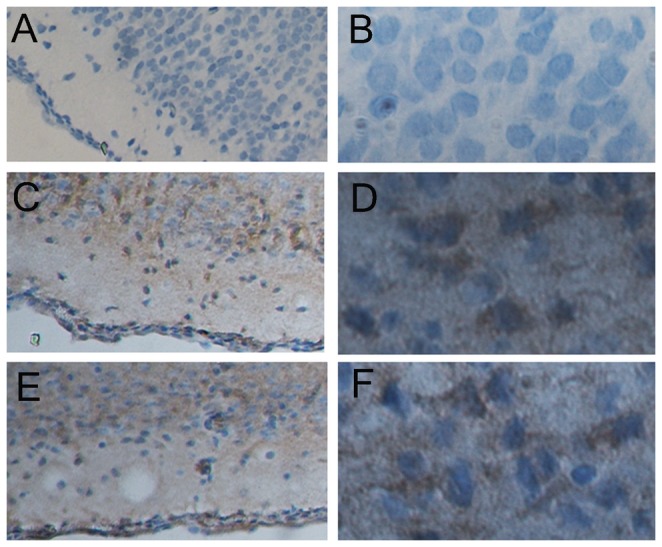
Current study explores the feasibility of using a non-surgical method of oxygen deprivation to create Hypoxic brain damage in neonatal rats for medical studies. 7-day-old Sprague Dowley (SD) rats were kept in a container with low oxygen level (8%) for 1.5h. A second group had bilateral cephalic artery ligation before the 1.5h-low oxygen treatment, a method similar to the popular Rice method, to expose the brain to both hypoxic and ischemic situations. Short term neural functions and brain water weights were evaluated 1 day after the hypoxic treatment. Brain pathology and histology were also examined at 1 day and 3 days after the hypoxic treatment. Both groups showed impaired neural functions and increased brain water weight compared to the controls. Histology studies also revealed injuries in the subcortex, hippocampus and lateral ventricle in the brains from both groups. There is no significant difference in the degree of brain damages observed in the two groups. Our work demonstrated that oxygen deprivation alone is sufficient to cause brain damages similar to those seen in Hypoxic-ischemic brain disease (HIBD). Because this method avoids the invasive surgical procedure and therefore reduces the stress and mortality of laboratory animals during the experiment, we recommend it to be the favorable method for creating rat models for HIBD studies.
当前的研究探索了使用非手术性缺氧方法在新生大鼠中造成缺氧性脑损伤以用于医学研究的可行性。将7日龄的斯普拉格-道利(SD)大鼠置于低氧水平(8%)的容器中1.5小时。第二组在进行1.5小时的低氧处理前进行双侧头动脉结扎,这是一种类似于常用的赖斯方法的手段,以使大脑暴露于缺氧和缺血的环境中。在缺氧处理1天后评估短期神经功能和脑水重量。在缺氧处理1天和3天后还检查了脑病理学和组织学情况。与对照组相比,两组均显示神经功能受损且脑水重量增加。组织学研究还揭示两组大鼠大脑的皮质下、海马体和侧脑室均有损伤。两组观察到的脑损伤程度无显著差异。我们的工作表明,单纯缺氧就足以导致与缺氧缺血性脑病(HIBD)中所见类似的脑损伤。由于该方法避免了侵入性手术操作,因此降低了实验过程中实验动物的应激和死亡率,我们推荐将其作为创建用于HIBD研究的大鼠模型的理想方法。