Simon Jacob C, Chan Kenneth H, Darling Cynthia L, Fried Daniel
Department of Preventive and Restorative Dental Sciences, University of California, San Francisco.
Lasers Surg Med. 2014 Mar;46(3):203-15. doi: 10.1002/lsm.22216. Epub 2013 Dec 27.
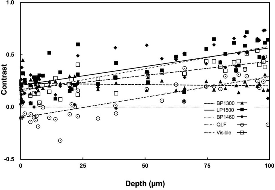
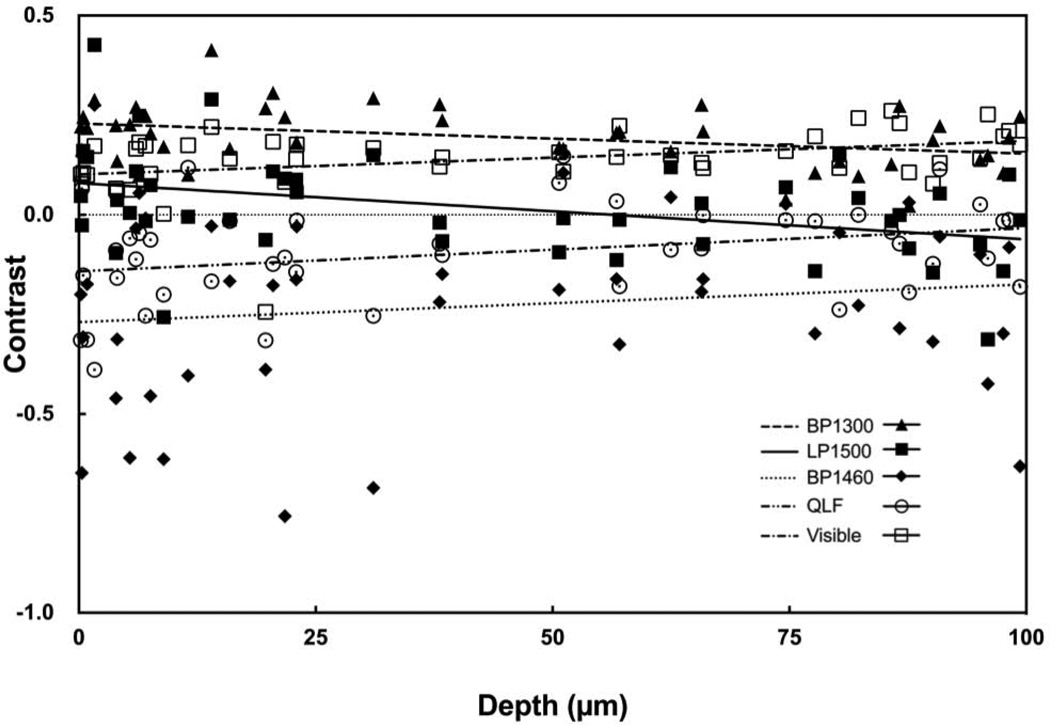
Early demineralization appears with high contrast at near-IR wavelengths due to a 10- to 20-fold difference in the magnitude of light scattering between sound and demineralized enamel. Water absorption in the near-IR has a significant effect on the lesion contrast and the highest contrast has been measured in spectral regions with higher water absorption. The purpose of this study was to determine how the lesion contrast changes with lesion severity and depth for different spectral regions in the near-IR and compare that range of contrast with visible reflectance and fluorescence.
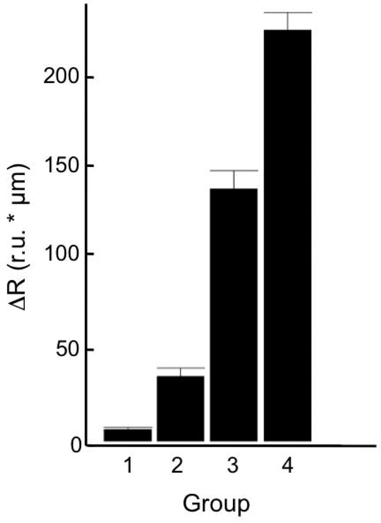
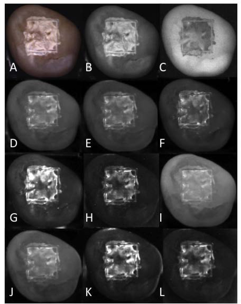
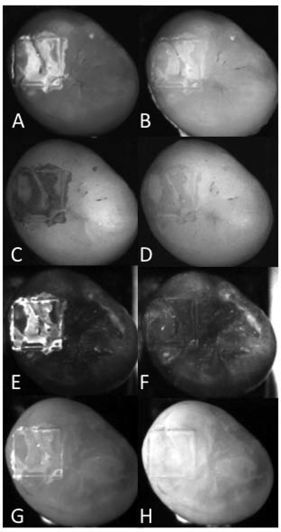
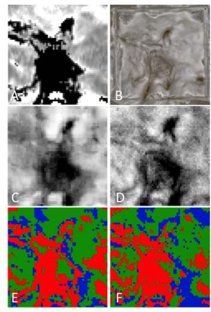
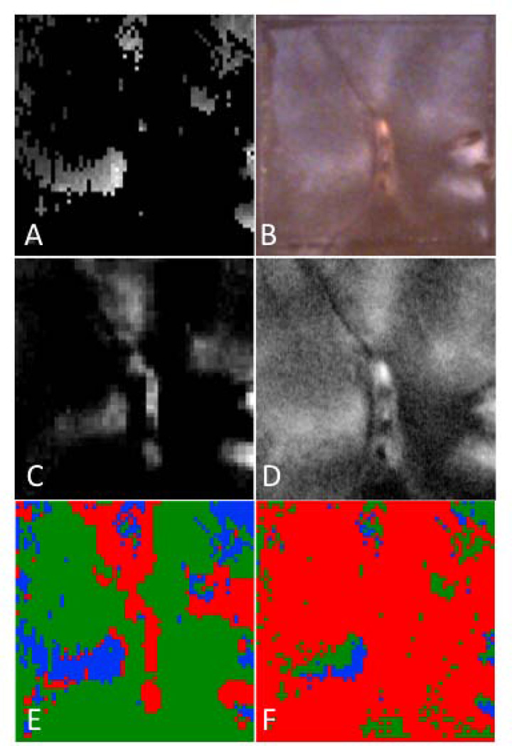
Forty-four human molars were used in this in vitro study. Teeth were painted with an acid-resistant varnish, leaving a 4 mm × 4 mm window on the occlusal surface of each tooth exposed for demineralization. Artificial lesions were produced in the unprotected windows after 12-48 hours exposure to a demineralizing solution at pH 4.5. Near-IR reflectance images were acquired over several near-IR spectral distributions, visible light reflectance, and fluorescence with 405-nm excitation and detection at wavelengths >500-nm. Crossed polarizers were used for reflectance measurements to reduce interference from specular reflectance. Cross polarization optical coherence tomography (CP-OCT) was used to non-destructively assess the depth and severity of demineralization in each sample window. Matching two-dimensional CP-OCT images of the lesion depth and integrated reflectivity were compared with the reflectance and fluorescence images to determine how accurately the variation in the lesion contrast represents the variation in the lesion severity.
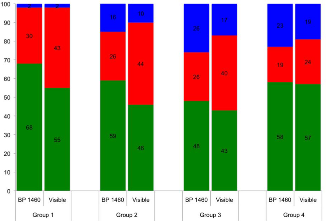
Artificial lesions appear more uniform on tooth surfaces exposed to an acid challenge at visible wavelengths than they do in the near-IR. Measurements of the lesion depth and severity using CP-OCT show that the lesion severity varies markedly across the sample windows and that the lesion contrast in the visible does not accurately reflect the large variation in the lesion severity. Reflectance measurements at certain near-IR wavelengths more accurately reflect variation in the depth and severity of the lesions.
The results of the study suggest that near-IR reflectance measurements at longer wavelengths coincident with higher water absorption are better suited for imaging early caries lesions.
由于完好牙釉质与脱矿牙釉质之间的光散射强度存在10至20倍的差异,早期脱矿在近红外波长下呈现出高对比度。近红外波段的水吸收对病变对比度有显著影响,且在水吸收较高的光谱区域测量到了最高的对比度。本研究的目的是确定在近红外的不同光谱区域中,病变对比度如何随病变严重程度和深度变化,并将该对比度范围与可见光反射率和荧光进行比较。
本体外研究使用了44颗人磨牙。用耐酸清漆涂抹牙齿,在每颗牙齿的咬合面上留出一个4毫米×4毫米的窗口暴露以进行脱矿。在pH值为4.5的脱矿溶液中暴露12至48小时后,在未受保护的窗口中产生人工病变。采集了多个近红外光谱分布、可见光反射率以及用405纳米激发并在波长>500纳米处检测的荧光的近红外反射图像。使用交叉偏振器进行反射率测量以减少镜面反射的干扰。使用交叉偏振光学相干断层扫描(CP-OCT)对每个样品窗口中的脱矿深度和严重程度进行无损评估。将病变深度与积分反射率的二维CP-OCT匹配图像与反射率和荧光图像进行比较,以确定病变对比度的变化在多大程度上准确反映了病变严重程度的变化。
在可见波长下,暴露于酸侵蚀的牙齿表面上的人工病变看起来比在近红外下更均匀。使用CP-OCT对病变深度和严重程度的测量表明,病变严重程度在样品窗口中变化显著,且可见光下的病变对比度不能准确反映病变严重程度的巨大差异。在某些近红外波长下的反射率测量更准确地反映了病变深度和严重程度的变化。
研究结果表明,与较高水吸收一致的较长波长下的近红外反射率测量更适合于早期龋损病变的成像。