Department of Radiology, Tokyo Metropolitan Geriatric Hospital and Institute of Gerontology 35-2 Sakaecho, Itabashi-ku, Tokyo, Japan ; Department of Nuclear Medicine, Saitama Medical University International Medical Center Saitama, Japan.
Integrative Brain Imaging Center, National Center of Neurology and Psychiatry Tokyo, Japan.
Brain Behav. 2013 Jul;3(4):487-93. doi: 10.1002/brb3.146. Epub 2013 Jun 30.
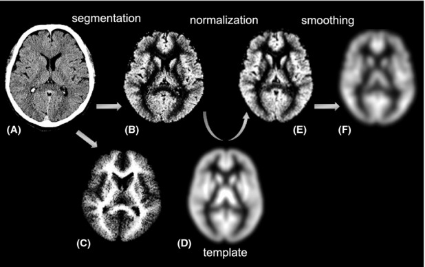
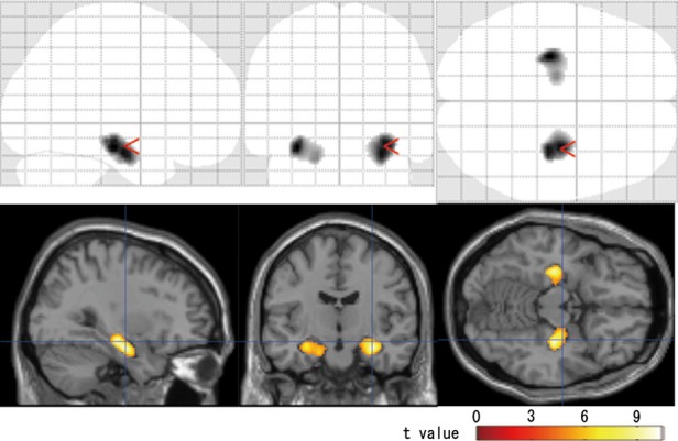
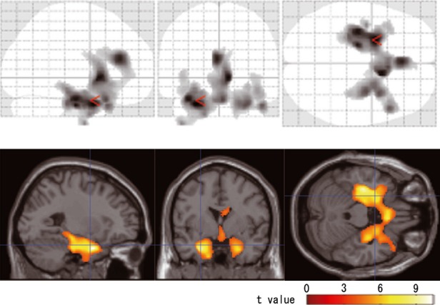
The voxel-based morphometry (VBM) technique using brain magnetic resonance imaging (MRI) objectively maps gray matter loss on a voxel-by-voxel basis after anatomic standardization. In patients with Alzheimer's disease (AD), reductions of gray matter volume, mainly in the medial temporal structures, have been reported; however, inhomogeneity and geometric distortion of the field intensity hampers the reproducibility of MRI. In the present study, we developed a novel computed tomography (CT)-based VBM method and used this technique to detect volume loss in AD patients as compared with normal controls. The results were compared with MRI-based VBM using the same subjects. Pittsburgh Compound B ((11)C-PIB) positron emission tomography (PET)/CT was performed and two experts in neuro-nuclear medicine judged whether regional amyloid β load was consistent with a diagnosis of AD. Before the injection of (11)C-PIB, high-quality CT scans were obtained using the same PET/CT equipment. MRI was performed within a mean interval of 25.1 ± 8.2 days before the PET/CT scan. Using statistical parametric mapping 8 (SPM8), the extracted gray matter images from CT and MRI were spatially normalized using a gray matter template and smoothed using a Gaussian kernel. Group comparisons were performed using SPM8 between five (11)C-PIB-positive patients with probable AD and seven (11)C-PIB-negative age-matched controls with normal cognition. Gray matter volumes in the bilateral medial temporal areas were reduced in the AD group as compared with the cognitively normal group in both CT-based VBM (in the left; P < 0.0001, cluster size 2776 and in the right; P < 0.0001, cluster size 630) and MRI-based VBM (in the left; P < 0.0001, cluster size 381 and in the right, P < 0.0001, cluster size 421). This newly developed CT-based VBM technique can detect significant atrophy in the entorhinal cortex in probable AD patients as previously reported using MRI-based VBM. However, CT-VBM was more sensitive and revealed larger areas of significant atrophy than MR-VBM.
基于体素的形态计量学 (VBM) 技术使用脑部磁共振成像 (MRI) 在解剖标准化后逐体素映射灰质损失。在阿尔茨海默病 (AD) 患者中,已经报道了灰质体积的减少,主要在中颞叶结构中;然而,场强的不均匀性和几何变形会阻碍 MRI 的可重复性。在本研究中,我们开发了一种新的基于计算机断层扫描 (CT) 的 VBM 方法,并使用该技术检测 AD 患者与正常对照之间的体积损失。将结果与使用相同受试者的基于 MRI 的 VBM 进行比较。进行匹兹堡复合物 B ((11)C-PIB) 正电子发射断层扫描 (PET)/CT,两位核医学专家判断区域淀粉样 β 负荷是否与 AD 诊断一致。在注射 (11)C-PIB 之前,使用相同的 PET/CT 设备获得高质量的 CT 扫描。MRI 在 PET/CT 扫描前平均间隔 25.1±8.2 天进行。使用统计参数映射 8 (SPM8),从 CT 和 MRI 中提取的灰质图像使用灰质模板进行空间归一化,并使用高斯核进行平滑处理。使用 SPM8 在 5 名(11)C-PIB 阳性可能 AD 患者和 7 名(11)C-PIB 阴性认知正常年龄匹配对照者之间进行组间比较。与认知正常组相比,AD 组双侧内侧颞叶区域的灰质体积在 CT 基于 VBM(左侧:P<0.0001,簇大小 2776,右侧:P<0.0001,簇大小 630)和 MRI 基于 VBM(左侧:P<0.0001,簇大小 381,右侧:P<0.0001,簇大小 421)中均减少。与之前使用 MRI 基于 VBM 报道的情况一样,这项新开发的 CT 基于 VBM 技术可以检测到可能的 AD 患者内嗅皮质的显著萎缩。然而,CT-VBM 更敏感,显示出比 MR-VBM 更大的显著萎缩区域。