• 文献检索
  • 文档翻译
  • 深度研究
  • 学术资讯
  • Suppr Zotero 插件Zotero 插件
  • 邀请有礼
  • 套餐&价格
  • 历史记录
应用&插件
Suppr Zotero 插件Zotero 插件浏览器插件Mac 客户端Windows 客户端微信小程序
定价
高级版会员购买积分包购买API积分包
服务
文献检索文档翻译深度研究API 文档MCP 服务
关于我们
关于 Suppr公司介绍联系我们用户协议隐私条款
关注我们

Suppr 超能文献

核心技术专利:CN118964589B侵权必究
粤ICP备2023148730 号-1Suppr @ 2026

文献检索

告别复杂PubMed语法,用中文像聊天一样搜索,搜遍4000万医学文献。AI智能推荐,让科研检索更轻松。

立即免费搜索

文件翻译

保留排版,准确专业,支持PDF/Word/PPT等文件格式,支持 12+语言互译。

免费翻译文档

深度研究

AI帮你快速写综述,25分钟生成高质量综述,智能提取关键信息,辅助科研写作。

立即免费体验

高级别胶质瘤的放射治疗:当前标准与新概念、影像与放射治疗的创新以及新治疗方法

Radiotherapy of high-grade gliomas: current standards and new concepts, innovations in imaging and radiotherapy, and new therapeutic approaches.

作者信息

Dhermain Frederic

机构信息

Department of Radiation Oncology, Institut Gustave Roussy University Hospital, Villejuif 94805, France.

出版信息

Chin J Cancer. 2014 Jan;33(1):16-24. doi: 10.5732/cjc.013.10217.

DOI:10.5732/cjc.013.10217
PMID:24384237
原文链接:https://pmc.ncbi.nlm.nih.gov/articles/PMC3905086/
Abstract

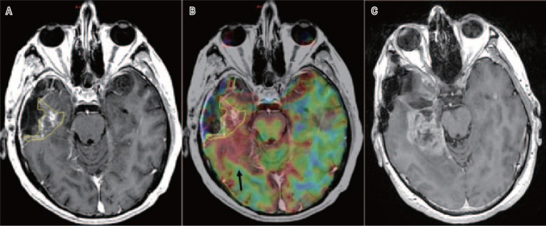
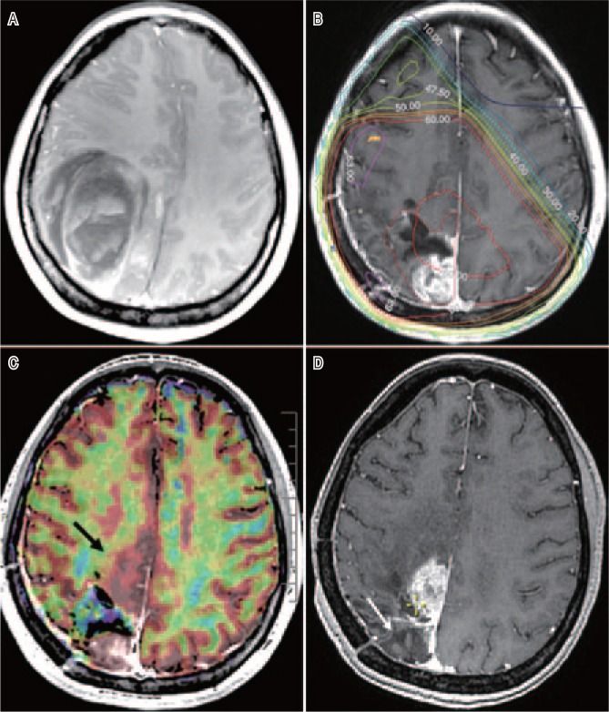
The current standards in radiotherapy of high-grade gliomas (HGG) are based on anatomic imaging techniques, usually computed tomography (CT) scanning and magnetic resonance imaging (MRI). The guidelines vary depending on whether the HGG is a histological grade 3 anaplastic glioma (AG) or a grade 4 glioblastoma multiforme (GBM). For AG, T2-weighted MRI sequences plus the region of contrast enhancement in T1 are considered for the delineation of the gross tumor volume (GTV), and an isotropic expansion of 15 to 20 mm is recommended for the clinical target volume (CTV). For GBM, the Radiation Therapy Oncology Group favors a two-step technique, with an initial phase (CTV1) including any T2 hyperintensity area (edema) plus a 20 mm margin treated with up to 46 Gy in 23 fractions, followed by a reduction in CTV2 to the contrast enhancement region in T1 with an additional 25 mm margin. The European Organisation of Research and Treatment of Cancer recommends a single-phase technique with a unique GTV, which comprises the T1 contrast enhancement region plus a margin of 20 to 30 mm. A total dose of 60 Gy in 30 fractions is usually delivered for GBM, and a dose of 59.4 Gy in 33 fractions is typically given for AG. As more than 85% of HGGs recur in field, dose-escalation studies have shown that 70 to 75 Gy can be delivered in 6 weeks with relevant toxicities developing in <10% of the patients. However, the only randomized dose-escalation trial, in which the boost dose was guided by conventional MRI, did not show any survival advantage of this treatment over the reference arm. HGGs are amongst the most infiltrative and heterogeneous tumors, and it was hypothesized that the most highly aggressive areas were missed; thus, better visualization of these high-risk regions for radiation boost could decrease the recurrence rate. Innovations in imaging and linear accelerators (LINAC) could help deliver the right doses of radiation to the right subvolumes according to the dose-painting concept. Advanced imaging techniques provide functional information on cellular density (diffusion MRI), angiogenesis (perfusion MRI), metabolic activity and cellular proliferation [positron emission tomography (PET) and magnetic resonance spectroscopy (MRS)]. All of these non-invasive techniques demonstrated good association between the images and histology, with up to 40% of HGGs functionally presenting a high activity within the non-contrast-enhanced areas in T1. New LINAC technologies, such as intensity-modulated and stereotactic radiotherapy, help to deliver a simultaneous integrated boost (SIB) > 60 Gy. Trials delivering a SIB into a biological GTV showed the feasibility of this treatment, but the final results, in terms of clinical benefits for HGG patients, are still pending. Many issues have been identified: the variety of MRI and PET machines (and amino-acid tracers), the heterogeneity of the protocols used for image acquisition and post-treatment, the geometric distortion and the unreliable algorithms for co-registration of brain anatomy with functional maps, and the semi-quiescent but highly invasive HGG cells. These issues could be solved by the homogenization of the protocols and software applications, the simultaneous acquisition of anatomic and functional images (PET-MRI machines), the combination of complementary imaging tools (perfusion and diffusion MRI), and the concomitant addition of some ad hoc targeted drugs against angiogenesis and invasiveness to chemoradiotherapy. The integration of these hybrid data will construct new synthetic metrics for fully individualized treatments.

https://cdn.ncbi.nlm.nih.gov/pmc/blobs/31b3/3905086/cac6e610304a/cjc-33-01-016-g002.jpg
https://cdn.ncbi.nlm.nih.gov/pmc/blobs/31b3/3905086/e4755ff41309/cjc-33-01-016-g001.jpg
https://cdn.ncbi.nlm.nih.gov/pmc/blobs/31b3/3905086/cac6e610304a/cjc-33-01-016-g002.jpg
https://cdn.ncbi.nlm.nih.gov/pmc/blobs/31b3/3905086/e4755ff41309/cjc-33-01-016-g001.jpg
https://cdn.ncbi.nlm.nih.gov/pmc/blobs/31b3/3905086/cac6e610304a/cjc-33-01-016-g002.jpg
摘要

高级别胶质瘤(HGG)放疗的当前标准基于解剖成像技术,通常是计算机断层扫描(CT)和磁共振成像(MRI)。指南因HGG是组织学3级间变性胶质瘤(AG)还是4级多形性胶质母细胞瘤(GBM)而异。对于AG,T2加权MRI序列加上T1加权成像中的对比增强区域用于勾勒大体肿瘤体积(GTV),建议临床靶体积(CTV)进行各向同性扩展15至20毫米。对于GBM,放射治疗肿瘤学组倾向于采用两步技术,初始阶段(CTV1)包括任何T2高信号区域(水肿)加上20毫米边缘,分23次给予高达46 Gy的剂量,随后将CTV2缩小至T1加权成像中的对比增强区域,并额外增加25毫米边缘。欧洲癌症研究与治疗组织推荐采用单相技术,即单一GTV,其包括T1加权成像对比增强区域加上20至30毫米的边缘。GBM通常分30次给予60 Gy的总剂量,AG通常分33次给予59.4 Gy的剂量。由于超过85%的HGG在野内复发,剂量递增研究表明,6周内可给予70至75 Gy的剂量,<10%的患者会出现相关毒性。然而,唯一一项随机剂量递增试验,其推量剂量由传统MRI引导,并未显示该治疗相对于对照臂有任何生存优势。HGG是最具浸润性和异质性的肿瘤之一,据推测最具侵袭性的区域被遗漏了;因此,更好地显示这些用于放射推量的高风险区域可能会降低复发率。成像和直线加速器(LINAC)的创新有助于根据剂量描绘概念将正确剂量的辐射传递到正确的子体积。先进的成像技术可提供有关细胞密度(扩散MRI)、血管生成(灌注MRI)、代谢活性和细胞增殖的功能信息[正电子发射断层扫描(PET)和磁共振波谱(MRS)]。所有这些非侵入性技术都显示出图像与组织学之间有良好的相关性,高达40%的HGG在T1加权成像的非对比增强区域内功能上呈现高活性。新的LINAC技术,如调强放疗和立体定向放疗,有助于给予同步整合推量(SIB)>60 Gy。将SIB应用于生物学GTV的试验表明了这种治疗的可行性,但就HGG患者的临床益处而言,最终结果仍有待确定。已发现许多问题:MRI和PET机器(以及氨基酸示踪剂)的多样性、用于图像采集和治疗后处理的方案的异质性、几何畸变以及用于脑解剖结构与功能图谱配准的不可靠算法,以及半静止但高度侵袭性的HGG细胞。这些问题可以通过使方案和软件应用同质化、同时采集解剖和功能图像(PET-MRI机器)、结合互补成像工具(灌注和扩散MRI)以及在放化疗中同时添加一些针对血管生成和侵袭性的特殊靶向药物来解决。这些混合数据的整合将构建用于完全个体化治疗的新综合指标。

相似文献

1
Radiotherapy of high-grade gliomas: current standards and new concepts, innovations in imaging and radiotherapy, and new therapeutic approaches.高级别胶质瘤的放射治疗:当前标准与新概念、影像与放射治疗的创新以及新治疗方法
Chin J Cancer. 2014 Jan;33(1):16-24. doi: 10.5732/cjc.013.10217.
2
Reirradiation of recurrent high-grade gliomas using amino acid PET (SPECT)/CT/MRI image fusion to determine gross tumor volume for stereotactic fractionated radiotherapy.使用氨基酸正电子发射断层扫描(单光子发射计算机断层扫描)/计算机断层扫描/磁共振成像图像融合技术对复发性高级别胶质瘤进行再照射,以确定立体定向分割放射治疗的大体肿瘤体积。
Int J Radiat Oncol Biol Phys. 2005 Oct 1;63(2):511-9. doi: 10.1016/j.ijrobp.2005.01.056.
3
Prospective Phase II Study of Radiotherapy Dose Escalation in Grade 4 Glioma Using Ga-Pentixafor PET Scan.采用 Ga-聚乙二醇化替莫唑胺 PET 扫描对 4 级胶质瘤进行放疗剂量递增的前瞻性 II 期研究。
Clin Oncol (R Coll Radiol). 2024 Sep;36(9):e294-e300. doi: 10.1016/j.clon.2024.04.011. Epub 2024 Apr 27.
4
Integration method of 3D MR spectroscopy into treatment planning system for glioblastoma IMRT dose painting with integrated simultaneous boost.将 3D MR 光谱学整合到治疗计划系统中,用于胶质母细胞瘤的 IMRT 剂量画中画同步强化治疗。
Radiat Oncol. 2013 Jan 2;8:1. doi: 10.1186/1748-717X-8-1.
5
Dose-painting multicenter phase III trial in newly diagnosed glioblastoma: the SPECTRO-GLIO trial comparing arm A standard radiochemotherapy to arm B radiochemotherapy with simultaneous integrated boost guided by MR spectroscopic imaging.新诊断胶质母细胞瘤的剂量描绘多中心 III 期试验:SPECTRO-GLIO 试验比较 A 组标准放化疗与 B 组放化疗联合 MR 波谱成像引导的同步综合增敏。
BMC Cancer. 2019 Feb 21;19(1):167. doi: 10.1186/s12885-019-5317-x.
6
Accelerated intensity-modulated radiotherapy plus temozolomide in patients with glioblastoma: a phase I dose-escalation study (ISIDE-BT-1).加速强度调制放疗联合替莫唑胺治疗胶质母细胞瘤患者的 I 期剂量递增研究(ISIDE-BT-1)。
Int J Clin Oncol. 2013 Oct;18(5):784-91. doi: 10.1007/s10147-012-0462-0. Epub 2012 Aug 15.
7
Efficacy of moderately hypofractionated simultaneous integrated boost intensity-modulated radiotherapy combined with temozolomide for the postoperative treatment of glioblastoma multiforme: a single-institution experience.适形调强同步推量放疗联合同步替莫唑胺化疗治疗多形性胶质母细胞瘤术后的疗效:单中心经验
Radiat Oncol. 2019 Jun 13;14(1):104. doi: 10.1186/s13014-019-1305-1.
8
Patterns of failure and comparison of different target volume delineations in patients with glioblastoma treated with conformal radiotherapy plus concomitant and adjuvant temozolomide.模式的失败和比较不同的目标体积描绘在患者胶质母细胞瘤治疗与适形放疗加上同期和辅助替莫唑胺。
Radiother Oncol. 2010 Dec;97(3):377-81. doi: 10.1016/j.radonc.2010.08.020. Epub 2010 Sep 18.
9
A phase I dose-escalation study (ISIDE-BT-1) of accelerated IMRT with temozolomide in patients with glioblastoma.一项关于替莫唑胺加速调强放疗治疗胶质母细胞瘤患者的 I 期剂量递增研究(ISIDE-BT-1)。
Int J Radiat Oncol Biol Phys. 2010 May 1;77(1):92-7. doi: 10.1016/j.ijrobp.2009.04.064. Epub 2009 Aug 14.
10
Single-arm phase II study of conformal radiation therapy and temozolomide plus fractionated stereotactic conformal boost in high-grade gliomas: final report.单臂二期研究:适形放疗联合替莫唑胺和分次立体定向适形推量治疗高级别胶质瘤:最终报告。
Strahlenther Onkol. 2010 Oct;186(10):558-64. doi: 10.1007/s00066-010-2101-x. Epub 2010 Sep 30.

引用本文的文献

1
A review of proton beam therapy's role in glioma management.质子束治疗在胶质瘤管理中的作用综述。
Medicine (Baltimore). 2025 Jul 4;104(27):e43071. doi: 10.1097/MD.0000000000043071.
2
Decitabine promotes the differentiation of poorly differentiated gastric cancer cells and enhances the sensitivity of NK cell cytotoxicity via TNF-α.地西他滨促进低分化胃癌细胞的分化,并通过肿瘤坏死因子-α增强自然杀伤细胞的细胞毒性敏感性。
Sci Rep. 2025 Apr 16;15(1):13119. doi: 10.1038/s41598-025-95741-0.
3
Recent Update on PET/CT Radiotracers for Imaging Cerebral Glioma.

本文引用的文献

1
Statistical analysis of multi-b factor diffusion weighted images can help distinguish between vasogenic and tumor-infiltrated edema.多b因子扩散加权图像的统计分析有助于区分血管源性水肿和肿瘤浸润性水肿。
J Magn Reson Imaging. 2014 Sep;40(3):622-9. doi: 10.1002/jmri.24399. Epub 2013 Nov 4.
2
Analysis of FET-PET imaging for target volume definition in patients with gliomas treated with conformal radiotherapy.分析适形放疗治疗脑胶质瘤患者时 FET-PET 成像在靶区勾画中的作用。
Radiother Oncol. 2013 Dec;109(3):487-92. doi: 10.1016/j.radonc.2013.06.043. Epub 2013 Aug 13.
3
RTOG 0913: a phase 1 study of daily everolimus (RAD001) in combination with radiation therapy and temozolomide in patients with newly diagnosed glioblastoma.
用于脑胶质瘤成像的PET/CT放射性示踪剂的最新进展
Nucl Med Mol Imaging. 2024 Jun;58(4):237-245. doi: 10.1007/s13139-024-00847-4. Epub 2024 Feb 21.
4
Exploring the Impact of Irradiation on Glioblastoma Blood-Brain-Barrier Permeability: Insights from Dynamic-Contrast-Enhanced-MRI and Histological Analysis.探索辐照对胶质母细胞瘤血脑屏障通透性的影响:来自动态对比增强磁共振成像和组织学分析的见解
Biomedicines. 2024 May 14;12(5):1091. doi: 10.3390/biomedicines12051091.
5
Radiomics for residual tumour detection and prognosis in newly diagnosed glioblastoma based on postoperative [C] methionine PET and T1c-w MRI.基于术后[C]蛋氨酸 PET 和 T1c-w MRI 的新诊断胶质母细胞瘤中残留肿瘤的检测和预后的影像组学
Sci Rep. 2024 Feb 25;14(1):4576. doi: 10.1038/s41598-024-55092-8.
6
Relationship between apparent diffusion coefficient and survival as a function of distance from gross tumor volume on radiation planning MRI in newly diagnosed glioblastoma.初诊胶质母细胞瘤放疗计划 MRI 上表观弥散系数与肿瘤全容积边缘距离与生存的关系
J Neurooncol. 2023 Sep;164(3):597-605. doi: 10.1007/s11060-023-04440-1. Epub 2023 Sep 14.
7
Understanding radiation response and cell cycle variation in brain tumour cells using Raman spectroscopy.使用拉曼光谱技术了解脑肿瘤细胞的辐射反应和细胞周期变化。
Analyst. 2023 May 30;148(11):2594-2608. doi: 10.1039/d3an00121k.
8
Stereotactic Magnetic Resonance-Guided Adaptive and Non-Adaptive Radiotherapy on Combination MR-Linear Accelerators: Current Practice and Future Directions.基于磁共振线性加速器组合的立体定向磁共振引导自适应与非自适应放射治疗:当前实践与未来方向。
Cancers (Basel). 2023 Mar 30;15(7):2081. doi: 10.3390/cancers15072081.
9
Evaluating the Utilization of Ethylenediaminetetraacetic Acid as a Treatment Supplement for Gliomas.评估乙二胺四乙酸作为胶质瘤治疗补充剂的应用情况。
Cureus. 2022 Nov 17;14(11):e31617. doi: 10.7759/cureus.31617. eCollection 2022 Nov.
10
A new approach to delineating clinical target volume for radiotherapy of glioblastoma: A phase II trial.一种用于胶质母细胞瘤放射治疗临床靶区勾画的新方法:一项II期试验。
Front Oncol. 2022 Oct 19;12:931436. doi: 10.3389/fonc.2022.931436. eCollection 2022.
RTOG 0913:每日依维莫司(RAD001)联合放疗和替莫唑胺治疗新诊断胶质母细胞瘤患者的 1 期研究。
Int J Radiat Oncol Biol Phys. 2013 Aug 1;86(5):880-4. doi: 10.1016/j.ijrobp.2013.04.036. Epub 2013 May 29.
4
Integral dose delivered to normal brain with conventional intensity-modulated radiotherapy (IMRT) and helical tomotherapy IMRT during partial brain radiotherapy for high-grade gliomas with and without selective sparing of the hippocampus, limbic circuit and neural stem cell compartment.在对高级别胶质瘤进行部分脑放疗时,采用传统调强放疗(IMRT)和螺旋断层放疗IMRT,在有或没有选择性保留海马体、边缘回路和神经干细胞区室的情况下,正常脑所接受的积分剂量。
J Med Imaging Radiat Oncol. 2013 Jun;57(3):378-83. doi: 10.1111/1754-9485.12048. Epub 2013 Apr 7.
5
The potential for an enhanced role for MRI in radiation-therapy treatment planning.磁共振成像(MRI)在放射治疗治疗计划中发挥更大作用的潜力。
Technol Cancer Res Treat. 2013 Oct;12(5):429-46. doi: 10.7785/tcrt.2012.500342. Epub 2013 Apr 24.
6
Prognostic vascular imaging biomarkers in high-grade gliomas: tumor permeability as an adjunct to blood volume estimates.高级别胶质瘤的预后血管成像生物标志物:肿瘤通透性作为血容量估计的辅助手段。
Acad Radiol. 2013 Apr;20(4):478-85. doi: 10.1016/j.acra.2012.11.011.
7
Biopsy validation of 18F-DOPA PET and biodistribution in gliomas for neurosurgical planning and radiotherapy target delineation: results of a prospective pilot study.18F-DOPA PET 与生物分布活检验证用于神经外科规划和放疗靶区勾画:一项前瞻性初步研究结果。
Neuro Oncol. 2013 Aug;15(8):1058-67. doi: 10.1093/neuonc/not002. Epub 2013 Mar 3.
8
Response classification based on a minimal model of glioblastoma growth is prognostic for clinical outcomes and distinguishes progression from pseudoprogression.基于胶质母细胞瘤生长的最小模型的反应分类对临床结局具有预后价值,并可区分进展和假性进展。
Cancer Res. 2013 May 15;73(10):2976-86. doi: 10.1158/0008-5472.CAN-12-3588. Epub 2013 Feb 11.
9
Advances in multimodal neuroimaging: hybrid MR-PET and MR-PET-EEG at 3 T and 9.4 T.多模态神经影像学进展:3T 和 9.4T 下的混合 MR-PET 和 MR-PET-EEG。
J Magn Reson. 2013 Apr;229:101-15. doi: 10.1016/j.jmr.2012.11.027. Epub 2012 Dec 10.
10
Integration method of 3D MR spectroscopy into treatment planning system for glioblastoma IMRT dose painting with integrated simultaneous boost.将 3D MR 光谱学整合到治疗计划系统中,用于胶质母细胞瘤的 IMRT 剂量画中画同步强化治疗。
Radiat Oncol. 2013 Jan 2;8:1. doi: 10.1186/1748-717X-8-1.