• 文献检索
  • 文档翻译
  • 深度研究
  • 学术资讯
  • Suppr Zotero 插件Zotero 插件
  • 邀请有礼
  • 套餐&价格
  • 历史记录
应用&插件
Suppr Zotero 插件Zotero 插件浏览器插件Mac 客户端Windows 客户端微信小程序
定价
高级版会员购买积分包购买API积分包
服务
文献检索文档翻译深度研究API 文档MCP 服务
关于我们
关于 Suppr公司介绍联系我们用户协议隐私条款
关注我们

Suppr 超能文献

核心技术专利:CN118964589B侵权必究
粤ICP备2023148730 号-1Suppr @ 2026

文献检索

告别复杂PubMed语法,用中文像聊天一样搜索,搜遍4000万医学文献。AI智能推荐,让科研检索更轻松。

立即免费搜索

文件翻译

保留排版,准确专业,支持PDF/Word/PPT等文件格式,支持 12+语言互译。

免费翻译文档

深度研究

AI帮你快速写综述,25分钟生成高质量综述,智能提取关键信息,辅助科研写作。

立即免费体验

不同但相似的干细胞类型对造影剂摄取的变异性。

Variability in contrast agent uptake by different but similar stem cell types.

机构信息

Biomedical MRI/MoSAIC, Department of Imaging and Pathology, Biomedical Sciences Group, Katholieke Universiteit Leuven, Leuven, Belgium.

Biomedical MRI/MoSAIC, Department of Imaging and Pathology, Biomedical Sciences Group, Katholieke Universiteit Leuven, Leuven, Belgium ; Lab of Histology, Biomedical Research Institute, Hasselt University, Campus Diepenbeek, Agoralaan, Diepenbeek, Belgium.

出版信息

Int J Nanomedicine. 2013;8:4577-91. doi: 10.2147/IJN.S51588. Epub 2013 Nov 29.

DOI:10.2147/IJN.S51588
PMID:24399873
原文链接:https://pmc.ncbi.nlm.nih.gov/articles/PMC3876490/
Abstract

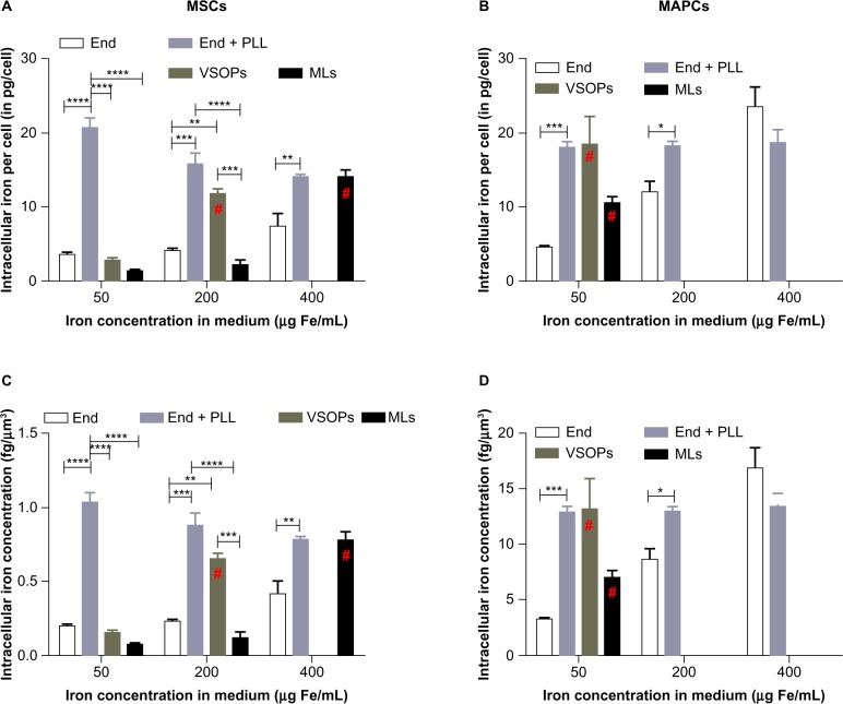
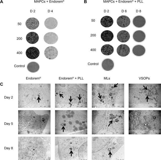
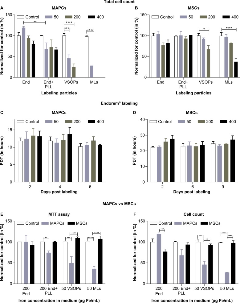
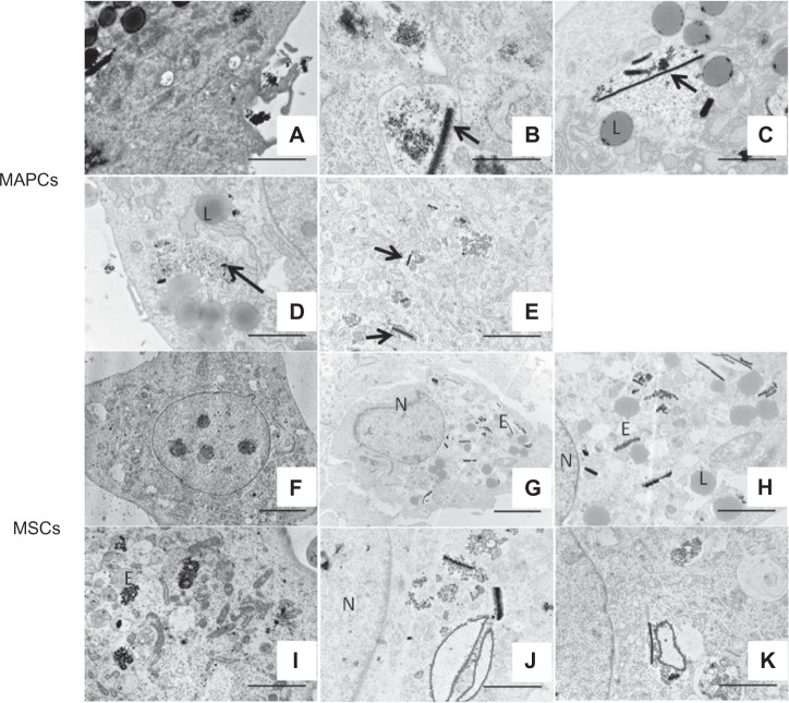
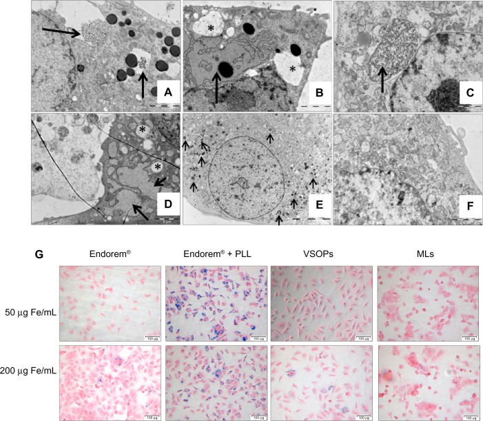
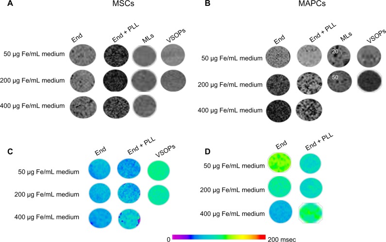
The need to track and evaluate the fate of transplanted cells is an important issue in regenerative medicine. In order to accomplish this, pre-labelling cells with magnetic resonance imaging (MRI) contrast agents is a well-established method. Uptake of MRI contrast agents by non-phagocytic stem cells, and factors such as cell homeostasis or the adverse effects of contrast agents on cell biology have been extensively studied, but in the context of nanoparticle (NP)-specific parameters. Here, we have studied three different types of NPs (Endorem®, magnetoliposomes [MLs], and citrate coated C-200) to label relatively larger, mesenchymal stem cells (MSCs) and, much smaller yet faster proliferating, multipotent adult progenitor cells (MAPCs). Both cell types are similar, as they are isolated from bone marrow and have substantial regenerative potential, which make them interesting candidates for comparative experiments. Using NPs with different surface coatings and sizes, we found that differences in the proliferative and morphological characteristics of the cells used in the study are mainly responsible for the fate of endocytosed iron, intracellular iron concentration, and cytotoxic responses. The quantitative analysis, using high-resolution electron microscopy images, demonstrated a strong relationship between cell volume/surface, uptake, and cytotoxicity. Interestingly, uptake and toxicity trends are reversed if intracellular concentrations, and not amounts, are considered. This indicates that more attention should be paid to cellular parameters such as cell size and proliferation rate in comparative cell-labeling studies.

摘要

追踪和评估移植细胞的命运是再生医学中的一个重要问题。为了实现这一目标,用磁共振成像(MRI)对比剂对细胞进行预标记是一种成熟的方法。非吞噬性干细胞对 MRI 对比剂的摄取,以及细胞内稳态或对比剂对细胞生物学的不良影响等因素,已经得到了广泛的研究,但都是在纳米颗粒(NP)的具体参数的背景下进行的。在这里,我们研究了三种不同类型的 NPs(钆塞酸二钠、磁脂体[MLs]和柠檬酸涂层 C-200)来标记相对较大的间充质干细胞(MSCs)和更小但增殖更快的多能成体祖细胞(MAPCs)。这两种细胞类型相似,因为它们都从骨髓中分离出来,具有很大的再生潜力,这使它们成为比较实验的有趣候选者。使用具有不同表面涂层和大小的 NPs,我们发现研究中使用的细胞的增殖和形态特征的差异主要是内吞铁的命运、细胞内铁浓度和细胞毒性反应的原因。使用高分辨率电子显微镜图像进行的定量分析表明,细胞体积/表面积、摄取和细胞毒性之间存在很强的关系。有趣的是,如果考虑细胞内浓度而不是数量,摄取和毒性趋势就会发生逆转。这表明,在比较细胞标记研究中,应该更加关注细胞大小和增殖率等细胞参数。

https://cdn.ncbi.nlm.nih.gov/pmc/blobs/e363/3876490/8bed27150d00/ijn-8-4577Fig6.jpg
https://cdn.ncbi.nlm.nih.gov/pmc/blobs/e363/3876490/2cd5070907cc/ijn-8-4577Fig1.jpg
https://cdn.ncbi.nlm.nih.gov/pmc/blobs/e363/3876490/1465a4503023/ijn-8-4577Fig2.jpg
https://cdn.ncbi.nlm.nih.gov/pmc/blobs/e363/3876490/946283a3844b/ijn-8-4577Fig3.jpg
https://cdn.ncbi.nlm.nih.gov/pmc/blobs/e363/3876490/3ac38f4a9ce0/ijn-8-4577Fig4.jpg
https://cdn.ncbi.nlm.nih.gov/pmc/blobs/e363/3876490/1b47857aa7f8/ijn-8-4577Fig5.jpg
https://cdn.ncbi.nlm.nih.gov/pmc/blobs/e363/3876490/8bed27150d00/ijn-8-4577Fig6.jpg

相似文献

1
Variability in contrast agent uptake by different but similar stem cell types.不同但相似的干细胞类型对造影剂摄取的变异性。
Int J Nanomedicine. 2013;8:4577-91. doi: 10.2147/IJN.S51588. Epub 2013 Nov 29.
2
Cytotoxic effects of iron oxide nanoparticles and implications for safety in cell labelling.氧化铁纳米颗粒的细胞毒性及其在细胞标记中的安全性意义。
Biomaterials. 2011 Jan;32(1):195-205. doi: 10.1016/j.biomaterials.2010.08.075. Epub 2010 Sep 22.
3
Co-precipitation of DEAE-dextran coated SPIONs: how synthesis conditions affect particle properties, stem cell labelling and MR contrast.DEAE-葡聚糖包被的超顺磁性氧化铁纳米颗粒的共沉淀:合成条件如何影响颗粒性质、干细胞标记和磁共振成像对比
Contrast Media Mol Imaging. 2016 Sep;11(5):362-370. doi: 10.1002/cmmi.1700. Epub 2016 Jun 30.
4
Magnetic labeling of non-phagocytic adherent cells with iron oxide nanoparticles: a comprehensive study.用氧化铁纳米颗粒对非吞噬性贴壁细胞进行磁性标记:一项综合研究。
Contrast Media Mol Imaging. 2008 Nov-Dec;3(6):223-32. doi: 10.1002/cmmi.256.
5
Heparin-coated superparamagnetic iron oxide for in vivo MR imaging of human MSCs.肝素涂层超顺磁性氧化铁在人骨髓间充质干细胞活体 MRI 成像中的应用。
Biomaterials. 2012 Jun;33(19):4861-71. doi: 10.1016/j.biomaterials.2012.03.035. Epub 2012 Apr 3.
6
Mesenchymal stem cell labeling and in vitro MR characterization at 1.5 T of new SPIO contrast agent: Molday ION Rhodamine-B™.新型超顺磁氧化铁对比剂 Molday ION Rhodamine-BTM 的 1.5T 磁共振成像特征及间质干细胞标记
Contrast Media Mol Imaging. 2011 Jan-Feb;6(1):7-18. doi: 10.1002/cmmi.396. Epub 2010 Aug 5.
7
Easy and Efficient Cell Tagging with Block Copolymer-Based Contrast Agents for Sensitive MRI Detection in Vivo.基于嵌段共聚物的造影剂实现简便高效的细胞标记用于体内灵敏磁共振成像检测
Cell Transplant. 2016 Oct;25(10):1787-1800. doi: 10.3727/096368916X691303.
8
Targeted Molecular Iron Oxide Contrast Agents for Imaging Atherosclerotic Plaque.靶向分子氧化铁对比剂用于动脉粥样硬化斑块成像。
Nanotheranostics. 2020 May 30;4(4):184-194. doi: 10.7150/ntno.44712. eCollection 2020.
9
Magnetic resonance imaging of ultrasmall superparamagnetic iron oxide-labeled exosomes from stem cells: a new method to obtain labeled exosomes.干细胞来源的超小型超顺磁性氧化铁标记外泌体的磁共振成像:一种获取标记外泌体的新方法。
Int J Nanomedicine. 2016 Jun 1;11:2481-90. doi: 10.2147/IJN.S104152. eCollection 2016.
10
Magnetoliposomes as magnetic resonance imaging contrast agents.磁性脂质体作为磁共振成像造影剂
Wiley Interdiscip Rev Nanomed Nanobiotechnol. 2011 Mar-Apr;3(2):197-211. doi: 10.1002/wnan.122. Epub 2011 Jan 28.

引用本文的文献

1
Cu-doped TiO nanoparticles improve local antitumor immune activation and optimize dendritic cell vaccine strategies.铜掺杂 TiO2 纳米颗粒改善局部抗肿瘤免疫激活并优化树突状细胞疫苗策略。
J Nanobiotechnology. 2023 Mar 13;21(1):87. doi: 10.1186/s12951-023-01844-z.
2
Superparamagnetic Iron Oxide Particles (VSOPs) Show Genotoxic Effects but No Functional Impact on Human Adipose Tissue-Derived Stromal Cells (ASCs).超顺磁性氧化铁颗粒(VSOPs)显示出遗传毒性作用,但对人脂肪组织来源的基质细胞(ASCs)无功能影响。
Materials (Basel). 2021 Jan 7;14(2):263. doi: 10.3390/ma14020263.
3
Development of Superparamagnetic Nanoparticles Coated with Polyacrylic Acid and Aluminum Hydroxide as an Efficient Contrast Agent for Multimodal Imaging.

本文引用的文献

1
Managing magnetic nanoparticle aggregation and cellular uptake: a precondition for efficient stem-cell differentiation and MRI tracking.管理磁性纳米颗粒聚集和细胞摄取:提高干细胞分化和 MRI 跟踪效率的前提。
Adv Healthc Mater. 2013 Feb;2(2):313-25. doi: 10.1002/adhm.201200294. Epub 2012 Nov 1.
2
Concise review: Bone marrow meets blastocyst: lessons from an unlikely encounter.简明回顾:骨髓遇见囊胚——一次意想不到的相遇带来的启示。
Stem Cells. 2013 Apr;31(4):620-6. doi: 10.1002/stem.1287.
3
Magnetic resonance imaging of human dental pulp stem cells in vitro and in vivo.
涂覆有聚丙烯酸和氢氧化铝的超顺磁性纳米颗粒作为多模态成像高效造影剂的研发
Nanomaterials (Basel). 2019 Nov 15;9(11):1626. doi: 10.3390/nano9111626.
4
Targeting tumor cells and neovascularization using RGD-functionalized magnetoliposomes.利用 RGD 功能化磁脂体靶向肿瘤细胞和新生血管。
Int J Nanomedicine. 2019 Jul 29;14:5911-5924. doi: 10.2147/IJN.S214041. eCollection 2019.
5
Improved Labeling of Pancreatic Islets Using Cationic Magnetoliposomes.使用阳离子磁脂质体改善胰岛标记
J Pers Med. 2018 Mar 12;8(1):12. doi: 10.3390/jpm8010012.
6
Intraarterial route increases the risk of cerebral lesions after mesenchymal cell administration in animal model of ischemia.动脉内给药途径会增加间充质细胞治疗缺血动物模型后脑损伤的风险。
Sci Rep. 2017 Jan 16;7:40758. doi: 10.1038/srep40758.
7
High content analysis at single cell level identifies different cellular responses dependent on nanomaterial concentrations.单细胞水平的高内涵分析可识别出依赖于纳米材料浓度的不同细胞反应。
Sci Rep. 2015 Sep 8;5:13890. doi: 10.1038/srep13890.
8
Nanoparticles and clinically applicable cell tracking.纳米颗粒与临床适用的细胞追踪
Br J Radiol. 2015 Oct;88(1054):20150375. doi: 10.1259/bjr.20150375. Epub 2015 Aug 7.
9
Cell type-specific response to high intracellular loading of polyacrylic acid-coated magnetic nanoparticles.细胞类型对聚丙烯酸包被磁性纳米颗粒高细胞内负载的特异性反应。
Int J Nanomedicine. 2015 Feb 18;10:1449-62. doi: 10.2147/IJN.S76134. eCollection 2015.
10
MRI-based identification of undifferentiated cells: looking at the two faces of Janus.基于磁共振成像的未分化细胞识别:审视双面雅努斯
Int J Nanomedicine. 2014 Feb 11;9:865-6. doi: 10.2147/IJN.S58674. eCollection 2014.
人牙髓干细胞的体外和体内磁共振成像。
Cell Transplant. 2013;22(10):1813-29. doi: 10.3727/096368912X657774. Epub 2012 Oct 8.
4
Human multipotent adult progenitor cells are nonimmunogenic and exert potent immunomodulatory effects on alloreactive T-cell responses.人多能成体祖细胞是免疫原性的,并对同种异体反应性 T 细胞反应发挥强大的免疫调节作用。
Cell Transplant. 2013;22(10):1915-28. doi: 10.3727/096368912X657369. Epub 2012 Oct 1.
5
Intravenous multipotent adult progenitor cell therapy after traumatic brain injury: modulation of the resident microglia population.创伤性脑损伤后的静脉注射多能成体祖细胞治疗:调节常驻小胶质细胞群体。
J Neuroinflammation. 2012 Sep 28;9:228. doi: 10.1186/1742-2094-9-228.
6
Therapeutic effects of hMAPC and hMSC transplantation after stroke in mice.hMAPC 和 hMSC 移植治疗小鼠卒中的疗效。
PLoS One. 2012;7(8):e43683. doi: 10.1371/journal.pone.0043683. Epub 2012 Aug 31.
7
MAPC culture conditions support the derivation of cells with nascent hypoblast features from bone marrow and blastocysts.MAPC培养条件支持从骨髓和囊胚中获得具有原始下胚层特征的细胞。
J Mol Cell Biol. 2012 Dec;4(6):423-6. doi: 10.1093/jmcb/mjs046. Epub 2012 Aug 9.
8
Combined transfection of the three transcriptional factors, PDX-1, NeuroD1, and MafA, causes differentiation of bone marrow mesenchymal stem cells into insulin-producing cells.三种转录因子PDX-1、NeuroD1和MafA的联合转染可使骨髓间充质干细胞分化为胰岛素分泌细胞。
Exp Diabetes Res. 2012;2012:672013. doi: 10.1155/2012/672013. Epub 2012 Jun 19.
9
Immune effects of mesenchymal stromal cells in experimental stroke.间质基质细胞在实验性中风中的免疫作用。
J Cereb Blood Flow Metab. 2012 Aug;32(8):1578-88. doi: 10.1038/jcbfm.2012.55. Epub 2012 May 2.
10
Limitations and caveats of magnetic cell labeling using transfection agent complexed iron oxide nanoparticles.使用转染试剂复合氧化铁纳米颗粒进行磁性细胞标记的局限性和注意事项。
Contrast Media Mol Imaging. 2012 Mar-Apr;7(2):140-52. doi: 10.1002/cmmi.472.