• 文献检索
  • 文档翻译
  • 深度研究
  • 学术资讯
  • Suppr Zotero 插件Zotero 插件
  • 邀请有礼
  • 套餐&价格
  • 历史记录
应用&插件
Suppr Zotero 插件Zotero 插件浏览器插件Mac 客户端Windows 客户端微信小程序
定价
高级版会员购买积分包购买API积分包
服务
文献检索文档翻译深度研究API 文档MCP 服务
关于我们
关于 Suppr公司介绍联系我们用户协议隐私条款
关注我们

Suppr 超能文献

核心技术专利:CN118964589B侵权必究
粤ICP备2023148730 号-1Suppr @ 2026

文献检索

告别复杂PubMed语法,用中文像聊天一样搜索,搜遍4000万医学文献。AI智能推荐,让科研检索更轻松。

立即免费搜索

文件翻译

保留排版,准确专业,支持PDF/Word/PPT等文件格式,支持 12+语言互译。

免费翻译文档

深度研究

AI帮你快速写综述,25分钟生成高质量综述,智能提取关键信息,辅助科研写作。

立即免费体验

Stargardt病视网膜斑点的功能分析

Functional Analysis of Retinal Flecks in Stargardt Disease.

作者信息

Verdina Tommaso, Tsang Stephen H, Greenstein Vivienne C, Zernant Jana, Sodi Andrea, Lima Luiz H, Chang Stanley, Allikmets Rando, Menchini Ugo

机构信息

Department of Specialized Surgical Sciences, Eye Clinic, University of Florence, Florence, Italy.

Department of Ophthalmology, Columbia University, New York, NY, USA ; Department of Pathology and Cell Biology, Columbia University, New York, NY, USA.

出版信息

J Clin Exp Ophthalmol. 2012 Jul 30;3. doi: 10.4172/2155-9570.1000233.

DOI:10.4172/2155-9570.1000233
PMID:24409374
原文链接:https://pmc.ncbi.nlm.nih.gov/articles/PMC3882688/
Abstract

PURPOSE

To evaluate visual function of flecked areas in a series of patients with Stargardt disease (STGD) and compare them with adjacent non flecked areas.

METHODS

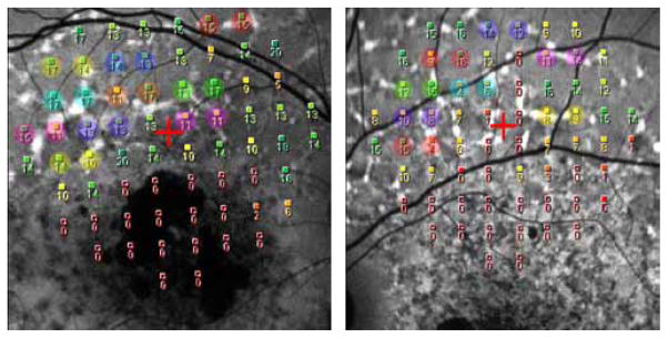
Twenty-seven patients with STGD, ABCA4 mutations and yellowish retinal flecks at fundus examination were recruited. Microperimetry with the Nidek MP-1 and fundus autofluorescence imaging (FAF) were performed in all the patients (27 eyes) while spectral-domain optical coherence tomography (SD-OCT) was performed in a subgroup of patients (20 eyes). Visual sensitivity (in dB) for each hyperfluorescent flecked area on FAF was compared with the value of the nearest adjacent non-flecked area in the MP-1 grid and at approximately the same distance from the fovea. Retinal structure in some of the flecked areas tested by microperimetry was analysed with SD-OCT. All patients were screened for mutations in the ABCA4 gene by APEX array and direct sequencing.

RESULTS

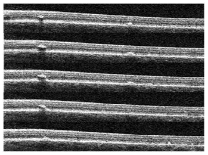
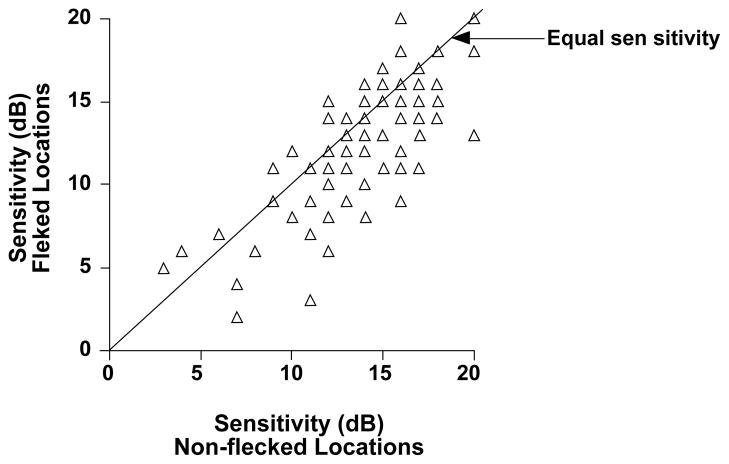
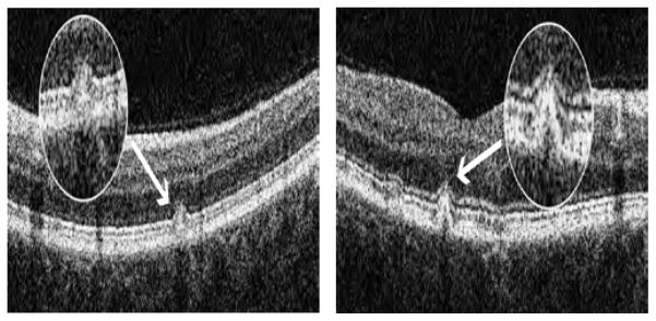
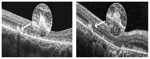
A total of 1836 locations (68 locations for each eye with the 10-2 program) were tested with the MP-1 and 97 corresponded to hyperautofluorescent flecks. A repeated measure, linear regression analysis was used to evaluate differences between visual sensitivity associated with the 97 flecked areas with those in the 97 neighbouring non-flecked areas. The difference was statistically significant (p<0.001) (flecked areas 12.89 +/- 3.86 dB vs. non-flecked areas 14.40 +/- 3.53 dB, respectively). SD-OCT in the flecked areas revealed the presence of hyperreflective dome-shaped lesions in the outer retina located at the level of the retinal pigment epithelium (RPE), with dislocation or disruption of the photoreceptor layer.

CONCLUSIONS

In STGD hyperfluorescent flecks on FAF are associated with decreased visual sensitivity compared to adjacent non-flecked areas and with an alteration of the photoreceptor layer on OCT. Flecks do not represent only a typical ophthalmoscopic feature but correspond, in some cases, to retinal damage that contributes to patients' visual loss.

摘要

目的

评估一系列斯塔加特病(STGD)患者斑点区域的视觉功能,并将其与相邻的无斑点区域进行比较。

方法

招募了27例经眼底检查确诊为STGD、ABCA4基因突变且有淡黄色视网膜斑点的患者。对所有患者(27只眼)进行了尼德克MP - 1微视野检查和眼底自发荧光成像(FAF),同时对部分患者(20只眼)进行了光谱域光学相干断层扫描(SD - OCT)。将FAF上每个高荧光斑点区域的视觉敏感度(以分贝为单位)与MP - 1网格中最近的相邻无斑点区域且距中央凹大致相同距离处的值进行比较。用SD - OCT分析了部分经微视野检查的斑点区域的视网膜结构。通过APEX阵列和直接测序对所有患者进行ABCA4基因突变筛查。

结果

用MP - 1共检测了1836个位置(采用10 - 2程序,每只眼68个位置),其中97个位置对应高自发荧光斑点。采用重复测量线性回归分析来评估与97个斑点区域相关的视觉敏感度与97个相邻无斑点区域的视觉敏感度之间的差异。差异具有统计学意义(p<0.001)(斑点区域分别为12.89±3.86分贝,无斑点区域为14.40±3.53分贝)。斑点区域的SD - OCT显示在视网膜色素上皮(RPE)水平的外层视网膜存在高反射穹顶状病变,伴有光感受器层的脱位或破坏。

结论

在STGD中,FAF上的高荧光斑点与相邻无斑点区域相比,视觉敏感度降低,且在OCT上光感受器层发生改变。斑点不仅是一种典型的检眼镜特征,在某些情况下还对应着导致患者视力丧失的视网膜损伤。

https://cdn.ncbi.nlm.nih.gov/pmc/blobs/ef85/3882688/0a7e1bf6b84b/nihms535866f5.jpg
https://cdn.ncbi.nlm.nih.gov/pmc/blobs/ef85/3882688/44df18383b15/nihms535866f1.jpg
https://cdn.ncbi.nlm.nih.gov/pmc/blobs/ef85/3882688/00b4f247c82f/nihms535866f2.jpg
https://cdn.ncbi.nlm.nih.gov/pmc/blobs/ef85/3882688/f7709e218480/nihms535866f3.jpg
https://cdn.ncbi.nlm.nih.gov/pmc/blobs/ef85/3882688/954a786e76c7/nihms535866f4.jpg
https://cdn.ncbi.nlm.nih.gov/pmc/blobs/ef85/3882688/0a7e1bf6b84b/nihms535866f5.jpg

相似文献

1
Functional Analysis of Retinal Flecks in Stargardt Disease.Stargardt病视网膜斑点的功能分析
J Clin Exp Ophthalmol. 2012 Jul 30;3. doi: 10.4172/2155-9570.1000233.
2
[Contribution of multimodal imaging in the various stages of Stargardt disease].[多模态成像在斯塔加特病各阶段的贡献]
J Fr Ophtalmol. 2017 Oct;40(8):666-675. doi: 10.1016/j.jfo.2017.05.008. Epub 2017 Sep 15.
3
Characterization of stargardt disease using polarization-sensitive optical coherence tomography and fundus autofluorescence imaging.应用偏振敏感光学相干断层扫描和眼底自发荧光成像技术对斯塔加特病进行特征描述。
Invest Ophthalmol Vis Sci. 2013 Sep 27;54(9):6416-25. doi: 10.1167/iovs.12-11550.
4
Photoreceptor cells as a source of fundus autofluorescence in recessive Stargardt disease.光感受器细胞作为隐性斯塔加特病眼底自发荧光的来源。
J Neurosci Res. 2019 Jan;97(1):98-106. doi: 10.1002/jnr.24252. Epub 2018 Apr 27.
5
Early-onset stargardt disease: phenotypic and genotypic characteristics.早发性斯塔加特病:表型和基因型特征。
Ophthalmology. 2015 Feb;122(2):335-44. doi: 10.1016/j.ophtha.2014.08.032. Epub 2014 Oct 17.
6
Analysis of retinal flecks in fundus flavimaculatus using high-definition spectral-domain optical coherence tomography.应用高清谱域光相干断层扫描分析眼底斑。
Am J Ophthalmol. 2010 Sep;150(3):330-7. doi: 10.1016/j.ajo.2010.04.001. Epub 2010 Jun 25.
7
Multimodal analysis of the Preferred Retinal Location and the Transition Zone in patients with Stargardt Disease.对斯塔加特病患者的视网膜优先位置和过渡区的多模态分析。
Graefes Arch Clin Exp Ophthalmol. 2017 Jul;255(7):1307-1317. doi: 10.1007/s00417-017-3637-6. Epub 2017 Apr 2.
8
Flecks in Recessive Stargardt Disease: Short-Wavelength Autofluorescence, Near-Infrared Autofluorescence, and Optical Coherence Tomography.隐性Stargardt病中的斑点:短波自发荧光、近红外自发荧光和光学相干断层扫描
Invest Ophthalmol Vis Sci. 2015 Jul;56(8):5029-39. doi: 10.1167/iovs.15-16763.
9
Analysis of retinal flecks in fundus flavimaculatus using optical coherence tomography.使用光学相干断层扫描分析黄斑黄点症中的视网膜斑点。
Br J Ophthalmol. 2006 Sep;90(9):1157-62. doi: 10.1136/bjo.2006.094136. Epub 2006 Jun 5.
10
Correlation of Morphology and Function of Flecks Using Short-Wave Fundus Autofluorescence and Microperimetry in Patients With Stargardt Disease.使用短波眼底自发荧光和微视野检查在 Stargardt 病患者中观察斑点的形态和功能相关性。
Transl Vis Sci Technol. 2021 Mar 1;10(3):18. doi: 10.1167/tvst.10.3.18.

引用本文的文献

1
Bisretinoid lipofuscin, fundus autofluorescence and retinal disease.双视黄醛脂褐质、眼底自发荧光与视网膜疾病。
Prog Retin Eye Res. 2025 Jul 8;108:101388. doi: 10.1016/j.preteyeres.2025.101388.
2
Quantifying the Progression of Stargardt Disease in Double-Null ABCA4 Carriers Using Fundus Autofluorescence Imaging.使用眼底自发荧光成像技术量化双等位基因ABCA4携带者中Stargardt病的进展情况。
Transl Vis Sci Technol. 2025 Mar 3;14(3):16. doi: 10.1167/tvst.14.3.16.
3
Multimodal Phenomap of Stargardt Disease Integrating Structural, Psychophysical, and Electrophysiologic Measures of Retinal Degeneration.

本文引用的文献

1
The inner segment/outer segment border seen on optical coherence tomography is less intense in patients with diminished cone function.在光学相干断层扫描中看到的内节/外节边界在 cone 功能下降的患者中强度降低。
Invest Ophthalmol Vis Sci. 2011 Dec 28;52(13):9703-9. doi: 10.1167/iovs.11-8650.
2
Analysis of the ABCA4 gene by next-generation sequencing.下一代测序分析 ABCA4 基因。
Invest Ophthalmol Vis Sci. 2011 Oct 31;52(11):8479-87. doi: 10.1167/iovs.11-8182.
3
Analysis of retinal flecks in fundus flavimaculatus using high-definition spectral-domain optical coherence tomography.
整合视网膜变性的结构、心理物理学和电生理学测量的Stargardt病多模态现象图谱
Ophthalmol Sci. 2023 May 9;4(1):100327. doi: 10.1016/j.xops.2023.100327. eCollection 2024 Jan-Feb.
4
The role of multimodal imaging and vision function testing in -related retinopathies and their relevance to future therapeutic interventions.多模态成像和视觉功能测试在相关视网膜病变中的作用及其与未来治疗干预的相关性。
Ther Adv Ophthalmol. 2021 Dec 19;13:25158414211056384. doi: 10.1177/25158414211056384. eCollection 2021 Jan-Dec.
5
Stargardt disease: Multimodal imaging: A review.斯特格病:多模态影像学:综述。
Clin Exp Ophthalmol. 2021 Jul;49(5):498-515. doi: 10.1111/ceo.13947. Epub 2021 Jun 1.
6
Correlation of Morphology and Function of Flecks Using Short-Wave Fundus Autofluorescence and Microperimetry in Patients With Stargardt Disease.使用短波眼底自发荧光和微视野检查在 Stargardt 病患者中观察斑点的形态和功能相关性。
Transl Vis Sci Technol. 2021 Mar 1;10(3):18. doi: 10.1167/tvst.10.3.18.
7
Occult inflammation detected by autofluorescence May Be the cause of idiopathic choroidal neovascularization.通过自体荧光检测到的隐匿性炎症可能是特发性脉络膜新生血管形成的原因。
Am J Ophthalmol Case Rep. 2020 Oct 17;20:100965. doi: 10.1016/j.ajoc.2020.100965. eCollection 2020 Dec.
8
Clinical spectrum, genetic complexity and therapeutic approaches for retinal disease caused by ABCA4 mutations.由 ABCA4 突变引起的视网膜疾病的临床谱、遗传复杂性和治疗方法。
Prog Retin Eye Res. 2020 Nov;79:100861. doi: 10.1016/j.preteyeres.2020.100861. Epub 2020 Apr 9.
9
Monitoring and Management of the Patient with Stargardt Disease.斯塔加特病患者的监测与管理
Clin Optom (Auckl). 2019 Nov 28;11:151-165. doi: 10.2147/OPTO.S226595. eCollection 2019.
10
EDI OCT evaluation of choroidal thickness in Stargardt disease.斯塔加特病脉络膜厚度的EDI OCT评估
PLoS One. 2018 Jan 5;13(1):e0190780. doi: 10.1371/journal.pone.0190780. eCollection 2018.
应用高清谱域光相干断层扫描分析眼底斑。
Am J Ophthalmol. 2010 Sep;150(3):330-7. doi: 10.1016/j.ajo.2010.04.001. Epub 2010 Jun 25.
4
In vivo visualization of photoreceptor layer and lipofuscin accumulation in stargardt's disease and fundus flavimaculatus by high resolution spectral-domain optical coherence tomography.利用高分辨率光谱域光学相干断层扫描技术对斯塔加特病和黄斑病变中光感受器层及脂褐素积聚进行体内可视化观察。
Clin Ophthalmol. 2009;3:693-9. doi: 10.2147/opth.s7894. Epub 2009 Dec 29.
5
Lipofuscin and autofluorescence metrics in progressive STGD.进行性Stargardt病中的脂褐素和自发荧光指标
Invest Ophthalmol Vis Sci. 2009 Aug;50(8):3907-14. doi: 10.1167/iovs.08-2448. Epub 2009 Apr 22.
6
Natural history of phenotypic changes in Stargardt macular dystrophy.斯塔加特黄斑营养不良表型变化的自然史。
Ophthalmic Genet. 2009 Jun;30(2):63-8. doi: 10.1080/13816810802695550.
7
A comparison of fundus autofluorescence and retinal structure in patients with Stargardt disease.Stargardt病患者的眼底自发荧光与视网膜结构比较
Invest Ophthalmol Vis Sci. 2009 Aug;50(8):3953-9. doi: 10.1167/iovs.08-2657. Epub 2009 Mar 25.
8
Novel mutations in of the ABCR gene in Italian patients with Stargardt disease.意大利斯塔加德特病患者 ABCR 基因的新型突变。
Eye (Lond). 2010 Jan;24(1):158-64. doi: 10.1038/eye.2009.35. Epub 2009 Mar 6.
9
Fixation stability measurement using the MP1 microperimeter.使用MP1微型视野计测量固视稳定性。
Retina. 2009 May;29(5):651-6. doi: 10.1097/IAE.0b013e318196bd65.
10
G1961E mutant allele in the Stargardt disease gene ABCA4 causes bull's eye maculopathy.斯塔加特病基因ABCA4中的G1961E突变等位基因导致靶心黄斑病变。
Exp Eye Res. 2009 Jun 15;89(1):16-24. doi: 10.1016/j.exer.2009.02.001. Epub 2009 Feb 13.