• 文献检索
  • 文档翻译
  • 深度研究
  • 学术资讯
  • Suppr Zotero 插件Zotero 插件
  • 邀请有礼
  • 套餐&价格
  • 历史记录
应用&插件
Suppr Zotero 插件Zotero 插件浏览器插件Mac 客户端Windows 客户端微信小程序
定价
高级版会员购买积分包购买API积分包
服务
文献检索文档翻译深度研究API 文档MCP 服务
关于我们
关于 Suppr公司介绍联系我们用户协议隐私条款
关注我们

Suppr 超能文献

核心技术专利:CN118964589B侵权必究
粤ICP备2023148730 号-1Suppr @ 2026

文献检索

告别复杂PubMed语法,用中文像聊天一样搜索,搜遍4000万医学文献。AI智能推荐,让科研检索更轻松。

立即免费搜索

文件翻译

保留排版,准确专业,支持PDF/Word/PPT等文件格式,支持 12+语言互译。

免费翻译文档

深度研究

AI帮你快速写综述,25分钟生成高质量综述,智能提取关键信息,辅助科研写作。

立即免费体验

斯塔加特病脉络膜厚度的EDI OCT评估

EDI OCT evaluation of choroidal thickness in Stargardt disease.

作者信息

Sodi Andrea, Bacherini Daniela, Lenzetti Chiara, Caporossi Orsola, Murro Vittoria, Mucciolo Dario Pasquale, Cipollini Francesca, Passerini Ilaria, Virgili Gianni, Rizzo Stanislao

机构信息

Department of Surgery and Translational Medicine, Eye Clinic, Careggi Teaching Hospital, Florence, Italy.

Department of Genetic Diagnosis, Careggi Teaching Hospital, Florence, Italy.

出版信息

PLoS One. 2018 Jan 5;13(1):e0190780. doi: 10.1371/journal.pone.0190780. eCollection 2018.

DOI:10.1371/journal.pone.0190780
PMID:29304098
原文链接:https://pmc.ncbi.nlm.nih.gov/articles/PMC5755895/
Abstract

PURPOSE

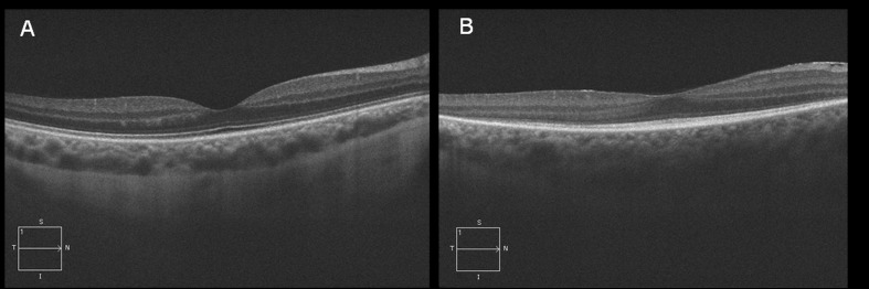
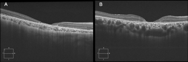
Choroidal thickness (CT) evaluation with EDI-OCT in Stargardt Disease (STGD), considering its possible association with some clinical features of the disease.

METHODS

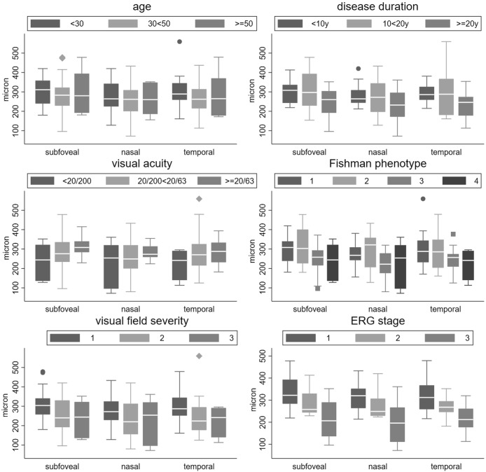
CT was evaluated in 41 STGD patients and in 70 controls. Measurements were performed in the subfoveal position and at 1000 μm nasally and temporally. CT average values in STGD and in the control group were first compared by means of Student's T test. Then, the possible association between CT and some clinical features was evaluated by means of linear regression analysis. Considered clinical parameters were: age, age on onset, duration of the disease, visual acuity, foveal thickness, Fishman clinical phenotype, visual field loss and ERG response.

RESULTS

Average CT was not significantly different between controls and STGD patients. In the STGD group the correlation between CT and age (r = 0.22, p = 0.033) and age of onset (r = 0.05, p = 0.424) was modest, while that of CT with disease duration (r = 0.30, p<0.001) was moderate. CT and foveal thickness were also significantly but modestly correlated (r = 0.15, p = 0.033).

CONCLUSION

In our series average CT is not significantly changed in STGD in comparison with the controls. Nevertheless a choroidal thinning may be identified in the more advanced stages of the disease.

摘要

目的

利用增强深度成像光学相干断层扫描(EDI-OCT)评估斯塔加特病(STGD)的脉络膜厚度(CT),并探讨其与该病某些临床特征的可能关联。

方法

对41例STGD患者和70例对照者进行CT评估。在黄斑中心凹下、鼻侧和颞侧1000μm处进行测量。首先通过学生t检验比较STGD组和对照组的CT平均值。然后,通过线性回归分析评估CT与某些临床特征之间的可能关联。考虑的临床参数包括:年龄、发病年龄、病程、视力、黄斑厚度、菲什曼临床表型、视野缺损和视网膜电图反应。

结果

对照组和STGD患者的平均CT无显著差异。在STGD组中,CT与年龄(r = 0.22,p = 0.033)和发病年龄(r = 0.05,p = 0.424)的相关性较弱,而CT与病程(r = 0.30,p<0.001)的相关性中等。CT与黄斑厚度也有显著但较弱的相关性(r = 0.15,p = 0.033)。

结论

在我们的系列研究中,与对照组相比,STGD患者的平均CT无显著变化。然而,在疾病的更晚期可能会发现脉络膜变薄。

https://cdn.ncbi.nlm.nih.gov/pmc/blobs/c0b7/5755895/a00120720af4/pone.0190780.g003.jpg
https://cdn.ncbi.nlm.nih.gov/pmc/blobs/c0b7/5755895/6550b43e6386/pone.0190780.g001.jpg
https://cdn.ncbi.nlm.nih.gov/pmc/blobs/c0b7/5755895/dfb2f10ea12b/pone.0190780.g002.jpg
https://cdn.ncbi.nlm.nih.gov/pmc/blobs/c0b7/5755895/a00120720af4/pone.0190780.g003.jpg
https://cdn.ncbi.nlm.nih.gov/pmc/blobs/c0b7/5755895/6550b43e6386/pone.0190780.g001.jpg
https://cdn.ncbi.nlm.nih.gov/pmc/blobs/c0b7/5755895/dfb2f10ea12b/pone.0190780.g002.jpg
https://cdn.ncbi.nlm.nih.gov/pmc/blobs/c0b7/5755895/a00120720af4/pone.0190780.g003.jpg

相似文献

1
EDI OCT evaluation of choroidal thickness in Stargardt disease.斯塔加特病脉络膜厚度的EDI OCT评估
PLoS One. 2018 Jan 5;13(1):e0190780. doi: 10.1371/journal.pone.0190780. eCollection 2018.
2
CHOROIDAL THICKNESS IN PATIENTS WITH STARGARDT DISEASE.斯特格病患者脉络膜厚度。
Retina. 2018 Mar;38(3):614-619. doi: 10.1097/IAE.0000000000001557.
3
Choroidal Thickness in Eyes With Central Geographic Atrophy Secondary to Stargardt Disease and Age-Related Macular Degeneration.继发于Stargardt病和年龄相关性黄斑变性的中心性地理萎缩眼中的脉络膜厚度
Ophthalmic Surg Lasers Imaging Retina. 2015 Sep;46(8):814-22. doi: 10.3928/23258160-20150909-05.
4
Characterization of stargardt disease using polarization-sensitive optical coherence tomography and fundus autofluorescence imaging.应用偏振敏感光学相干断层扫描和眼底自发荧光成像技术对斯塔加特病进行特征描述。
Invest Ophthalmol Vis Sci. 2013 Sep 27;54(9):6416-25. doi: 10.1167/iovs.12-11550.
5
Clinical and molecular characteristics of childhood-onset Stargardt disease.儿童期发病的斯塔加特病的临床和分子特征
Ophthalmology. 2015 Feb;122(2):326-34. doi: 10.1016/j.ophtha.2014.08.012. Epub 2014 Oct 12.
6
EDI-OCT evaluation of choroidal thickness in retinitis pigmentosa.视网膜色素变性中脉络膜厚度的光学相干断层扫描血管造影评估
Eur J Ophthalmol. 2018 Jan;28(1):52-57. doi: 10.5301/ejo.5000961.
7
Morphology and Vascular Layers of the Choroid in Stargardt Disease Analyzed Using Spectral-Domain Optical Coherence Tomography.使用光谱域光学相干断层扫描分析斯塔加特病脉络膜的形态学和血管层
Am J Ophthalmol. 2015 Dec;160(6):1276-1284.e1. doi: 10.1016/j.ajo.2015.08.025. Epub 2015 Aug 24.
8
Dark Atrophy: An Optical Coherence Tomography Angiography Study.暗萎缩:光学相干断层扫描血管造影研究。
Ophthalmology. 2016 Sep;123(9):1879-86. doi: 10.1016/j.ophtha.2016.05.041. Epub 2016 Jul 19.
9
Optical Coherence Tomography Angiography Findings in Stargardt Disease.斯塔加特病的光学相干断层扫描血管造影结果
PLoS One. 2017 Feb 2;12(2):e0170343. doi: 10.1371/journal.pone.0170343. eCollection 2017.
10
Correlation between photoreceptor layer integrity and visual function in patients with Stargardt disease: implications for gene therapy.Stargardt 病患者的光感受器层完整性与视觉功能的相关性:对基因治疗的启示。
Invest Ophthalmol Vis Sci. 2012 Jul 3;53(8):4409-15. doi: 10.1167/iovs.11-8201.

引用本文的文献

1
A deep learning model for diagnosis of inherited retinal diseases.一种用于诊断遗传性视网膜疾病的深度学习模型。
Sci Rep. 2025 Jul 2;15(1):22523. doi: 10.1038/s41598-025-04648-3.
2
Effects of exogenous retinoic acid on ocular parameters in Guinea pigs with form deprivation myopia.外源性视黄酸对形觉剥夺性近视豚鼠眼部参数的影响。
Front Cell Dev Biol. 2023 Mar 17;11:1160897. doi: 10.3389/fcell.2023.1160897. eCollection 2023.
3
Choroidal Caverns in Stargardt Disease.斯特格病中的脉络膜海绵状血管病变。

本文引用的文献

1
CHOROIDAL THICKNESS IN PATIENTS WITH STARGARDT DISEASE.斯特格病患者脉络膜厚度。
Retina. 2018 Mar;38(3):614-619. doi: 10.1097/IAE.0000000000001557.
2
Assessment of estimated retinal atrophy progression in Stargardt macular dystrophy using spectral-domain optical coherence tomography.使用频域光学相干断层扫描评估斯塔加特黄斑营养不良中视网膜萎缩的估计进展情况。
Br J Ophthalmol. 2016 Jul;100(7):956-962. doi: 10.1136/bjophthalmol-2015-307035. Epub 2015 Nov 14.
3
Choroidal Thickness in Eyes With Central Geographic Atrophy Secondary to Stargardt Disease and Age-Related Macular Degeneration.
Invest Ophthalmol Vis Sci. 2022 Feb 1;63(2):25. doi: 10.1167/iovs.63.2.25.
4
Localized Structural and Functional Deficits in a Nonhuman Primate Model of Outer Retinal Atrophy.局部结构和功能缺陷的非人类灵长类动物模型的外层视网膜萎缩。
Invest Ophthalmol Vis Sci. 2021 Oct 4;62(13):8. doi: 10.1167/iovs.62.13.8.
5
Choroidal Thickness in Different Types of Inherited Retinal Dystrophies.不同类型遗传性视网膜营养不良中的脉络膜厚度
J Ophthalmic Vis Res. 2020 Aug 6;15(3):351-361. doi: 10.18502/jovr.v15i3.7454. eCollection 2020 Jul-Sep.
继发于Stargardt病和年龄相关性黄斑变性的中心性地理萎缩眼中的脉络膜厚度
Ophthalmic Surg Lasers Imaging Retina. 2015 Sep;46(8):814-22. doi: 10.3928/23258160-20150909-05.
4
Morphology and Vascular Layers of the Choroid in Stargardt Disease Analyzed Using Spectral-Domain Optical Coherence Tomography.使用光谱域光学相干断层扫描分析斯塔加特病脉络膜的形态学和血管层
Am J Ophthalmol. 2015 Dec;160(6):1276-1284.e1. doi: 10.1016/j.ajo.2015.08.025. Epub 2015 Aug 24.
5
ET-1 Plasma Levels, Aqueous Flare, and Choroidal Thickness in Patients with Retinitis Pigmentosa.色素性视网膜炎患者的血浆内皮素-1水平、房水闪光及脉络膜厚度
J Ophthalmol. 2015;2015:292615. doi: 10.1155/2015/292615. Epub 2015 Jun 2.
6
Morpho-functional correlation of fundus autofluorescence in Stargardt disease.Stargardt病中眼底自发荧光的形态功能相关性
Br J Ophthalmol. 2015 Oct;99(10):1354-9. doi: 10.1136/bjophthalmol-2014-306237. Epub 2015 Apr 2.
7
Choroidal thickness in geographic atrophy secondary to age-related macular degeneration.年龄相关性黄斑变性继发地图样萎缩的脉络膜厚度。
Invest Ophthalmol Vis Sci. 2015 Jan 13;56(2):875-82. doi: 10.1167/iovs.14-14933.
8
Relationship of central choroidal thickness with age-related macular degeneration status.中心脉络膜厚度与年龄相关性黄斑变性状态的关系。
Am J Ophthalmol. 2015 Apr;159(4):617-26. doi: 10.1016/j.ajo.2014.12.010. Epub 2014 Dec 17.
9
Enhanced depth imaging-OCT of the choroid: a review of the current literature.脉络膜增强深度成像光学相干断层扫描:当前文献综述
Graefes Arch Clin Exp Ophthalmol. 2014 Dec;252(12):1871-83. doi: 10.1007/s00417-014-2840-y. Epub 2014 Nov 4.
10
Quantitative fundus autofluorescence in recessive Stargardt disease.隐性Stargardt病的定量眼底自发荧光
Invest Ophthalmol Vis Sci. 2014 May 1;55(5):2841-52. doi: 10.1167/iovs.13-13624.