Xiong Zhi, Thangavel Ramasamy, Kempuraj Duraisamy, Yang Evert, Zaheer Smita, Zaheer Asgar
Department of Neurology, Carver College of Medicine, University of Iowa Hospitals and Clinics, Iowa City, IA, USA.
Veterans Affairs Health Care System, Iowa City, IA, USA Department of Neurology, Carver College of Medicine, University of Iowa Hospitals and Clinics, Iowa City, IA, USA.
J Alzheimers Dis. 2014;40(2):297-308. doi: 10.3233/JAD-132081.
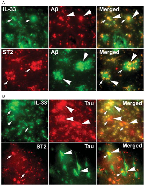
Inflammatory responses are increasingly implicated in the pathogenesis of neurodegenerative diseases such as in Alzheimer's disease (AD). Interleukin-33 (IL-33), a member of IL-1 family, is constitutively expressed in the central nervous system and thought to be an important mediator of glial cell response to neuropathological lesions. Proinflammatory molecules are highly expressed at the vicinity of amyloid plaques (APs) and neurofibrillary tangles (NFTs), the hallmarks of AD pathology. We have investigated the expression of IL-33 and ST2 in relation to APs and NFTs in human AD and non-AD control brains by immunohistochemistry. Sections from the entorhinal cortex, where APs and NFTs appear in early stages of AD, were used for immunohistochemistry. Mouse primary astrocytes were cultured and incubated with amyloid-β1-42 (Aβ1-42), component of plaque for 72 h and analyzed for the expression of IL-33 by flow cytometry. We found strong expression of IL-33 and ST2 in the vicinity of Aβ and AT8 labelled APs and NFTs respectively, and in the glial cells in AD brains when compared to non-AD control brains. IL-33 and ST2 positive cells were also significantly increased in AD brains when compared to non-AD brains. Flow cytometric analysis revealed incubation of mouse astrocytes with Aβ1-42 increased astrocytic IL-33 expression in vitro. These results suggest that IL-33, an alamin cytokine, may induce inflammatory molecule release from the glial cells and may play an important role in the pathogenesis of AD.
炎症反应越来越多地被认为与神经退行性疾病(如阿尔茨海默病,AD)的发病机制有关。白细胞介素-33(IL-33)是IL-1家族的一员,在中枢神经系统中组成性表达,被认为是胶质细胞对神经病理损伤反应的重要介质。促炎分子在淀粉样斑块(APs)和神经原纤维缠结(NFTs)附近高度表达,而APs和NFTs是AD病理的标志。我们通过免疫组织化学研究了人类AD和非AD对照脑中IL-33和ST2与APs和NFTs相关的表达情况。取自内嗅皮质的切片用于免疫组织化学,内嗅皮质是AD早期出现APs和NFTs的部位。培养小鼠原代星形胶质细胞,并用斑块成分β淀粉样蛋白1-42(Aβ1-42)孵育72小时,然后通过流式细胞术分析IL-33的表达。我们发现,与非AD对照脑相比,在AD脑中,IL-33和ST2分别在Aβ和AT8标记的APs和NFTs附近以及胶质细胞中有强烈表达。与非AD脑相比,AD脑中IL-33和ST2阳性细胞也显著增加。流式细胞术分析显示,用Aβ1-42孵育小鼠星形胶质细胞可在体外增加星形胶质细胞IL-33的表达。这些结果表明,IL-33作为一种警报素细胞因子,可能诱导胶质细胞释放炎症分子,并可能在AD的发病机制中起重要作用。