Gröger Adriane, Kolb Rupert, Schäfer Rita, Klose Uwe
Department of Diagnostic and Interventional Neuroradiology, Magnetic Resonance Research Group, University Hospital Tübingen, Tübingen, Germany.
PLoS One. 2014 Jan 8;9(1):e84081. doi: 10.1371/journal.pone.0084081. eCollection 2014.
Metabolic changes in the substantia nigra of patients with Parkinson's disease were previously investigated in different molecular-pathological examinations. The aim of our study was the in vivo measurement of these alterations using three-dimensional magnetic resonance spectroscopic imaging.
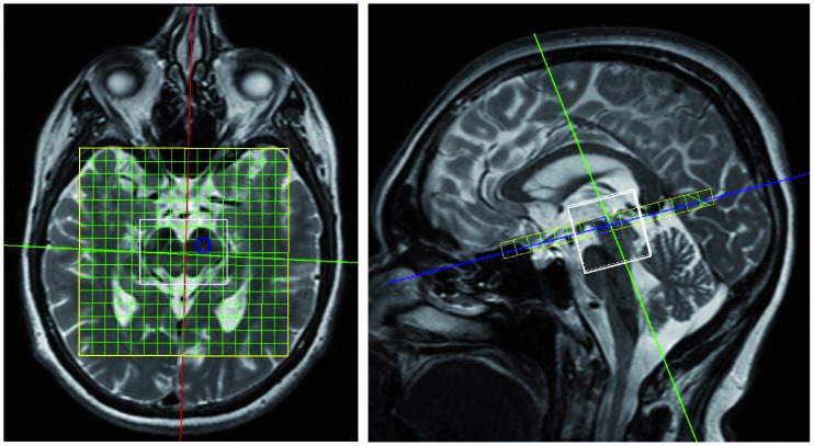
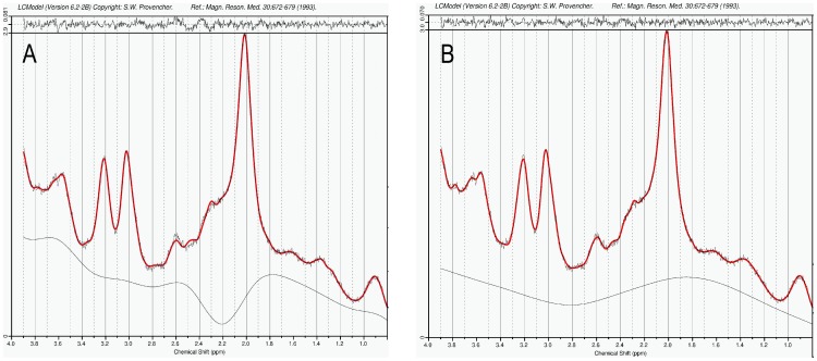
21 patients with Parkinson's disease and 24 controls were examined using magnetic resonance spectroscopic imaging at 3 Tesla. The spectra of rostral and caudal substantia nigra regions were analyzed using LCModel. For spectral fitting, an adjusted basis data set with pathology-specific metabolites and macromolecules was used to better reproduce the in vivo spectra. To assess differences between both groups more accurately, especially in metabolites at lower concentrations, group-averaged spectra were evaluated in addition to the analysis of individual data.
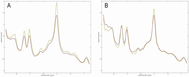
We found significantly decreased N-acetylaspartate, choline, creatine, myo-inositol, glutathione and dopamine concentrations in patients with Parkinson's disease compared to controls, whereas glutamine+glutamate, γ-aminobutyric acid, and homovanillic acid were slightly increased. According to anatomical features, clear differences in the biochemical profiles were found between rostral and caudal substantia nigra voxels in both groups.
Reduced N-acetylaspartate and dopamine concentrations result from progressive degeneration of dopamine-producing neurons within the substantia nigra pars compacta. Decreased creatine levels can be interpreted as impaired energy metabolism due to mitochondrial dysfunction. Lower glutathione concentrations might be a cause or consequence of oxidative stress. Furthermore, slightly increased glutamine+glutamate and γ-aminobutyric acid levels are expected based on post mortem data in Parkinson's disease. To the best of our knowledge, this is the first non-invasive confirmation of these metabolic changes.
先前在不同的分子病理学检查中对帕金森病患者黑质的代谢变化进行了研究。我们研究的目的是使用三维磁共振波谱成像对这些改变进行活体测量。
对21例帕金森病患者和24名对照者进行3特斯拉磁共振波谱成像检查。使用LCModel分析黑质头端和尾端区域的波谱。为进行波谱拟合,使用了包含病理学特异性代谢物和大分子的调整后基础数据集,以更好地重现活体波谱。为了更准确地评估两组之间的差异,特别是低浓度代谢物的差异,除了分析个体数据外,还对组平均波谱进行了评估。
我们发现,与对照组相比,帕金森病患者的N-乙酰天门冬氨酸、胆碱、肌酸、肌醇、谷胱甘肽和多巴胺浓度显著降低,而谷氨酰胺+谷氨酸、γ-氨基丁酸和高香草酸略有升高。根据解剖学特征,两组黑质头端和尾端体素的生化特征存在明显差异。
N-乙酰天门冬氨酸和多巴胺浓度降低是由于黑质致密部多巴胺能神经元的进行性退变所致。肌酸水平降低可解释为线粒体功能障碍导致能量代谢受损。谷胱甘肽浓度降低可能是氧化应激的原因或结果。此外,根据帕金森病的尸检数据,预计谷氨酰胺+谷氨酸和γ-氨基丁酸水平会略有升高。据我们所知,这是这些代谢变化的首次非侵入性证实。