Davis Anne E, Lewandowski Adam J, Holloway Cameron J, Ntusi Ntobeko A B, Banerjee Rajarshi, Nethononda Richard, Pitcher Alex, Francis Jane M, Myerson Saul G, Leeson Paul, Donovan Tim, Neubauer Stefan, Rider Oliver J
Radcliffe Department of Medicine, Division of Cardiovascular Medicine, John Radcliffe Hospital, Oxford OX3 9DU, UK.
J Cardiovasc Magn Reson. 2014 Jan 21;16(1):9. doi: 10.1186/1532-429X-16-9.
Cardiovascular magnetic resonance (CMR) is regarded as the gold standard for clinical assessment of the aorta, but normal dimensions are usually referenced to echocardiographic and computed tomography data and no large CMR normal reference range exists. As a result we aimed to 1) produce a normal CMR reference range of aortic diameters and 2) investigate the relationship between regional aortic size and body surface area (BSA) in a large group of healthy subjects with no vascular risk factors.
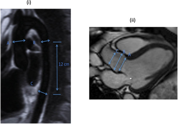
447 subjects (208 male, aged 19-70 years) without identifiable cardiac risk factors (BMI range 15.7-52.6 kg/m2) underwent CMR at 1.5 T to determine aortic diameter at three levels: the ascending aorta (Ao) and proximal descending aorta (PDA) at the level of the pulmonary artery, and the abdominal aorta (DDA), at a level 12 cm distal to the PDA. In addition, 201 of these subjects had aortic root imaging, allowing for measurements at the level of the aortic valve annulus (AV), aortic sinuses and sinotubular junction (STJ).
Normal diameters (mean ±2 SD) were; AV annulus male(♂) 24.4 ± 5.4, female (♀) 21.0 ± 3.6 mm, aortic sinus♂ 32.4 ± 7.7, ♀27.6 ± 5.8 mm, ST-junction ♂25.0 ± 7.4, ♀21.8 ± 5.4 mm, Ao ♂26.7 ± 7.7, ♀25.5 ± 7.4 mm, PDA ♂20.6 ± 5.6, +18.9 ± 4.0 mm, DDA ♂17.6 ± 5.1, ♀16.4 ± 4.0 mm. Aortic root and thoracic aortic diameters increased at all levels measured with BSA. No gender difference was seen in the degree of dilatation with increasing BSA (p>0.5 for all analyses).
Across both genders, increasing body size is characterized by a modest degree of aortic dilatation, even in the absence of traditional cardiovascular risk factors.
心血管磁共振成像(CMR)被视为主动脉临床评估的金标准,但正常尺寸通常参考超声心动图和计算机断层扫描数据,且不存在大型CMR正常参考范围。因此,我们旨在:1)得出主动脉直径的正常CMR参考范围;2)在一大群无血管危险因素的健康受试者中研究局部主动脉大小与体表面积(BSA)之间的关系。
447名无明确心脏危险因素(BMI范围为15.7 - 52.6 kg/m²)的受试者(208名男性,年龄19 - 70岁)接受了1.5T的CMR检查,以确定三个水平的主动脉直径:肺动脉水平的升主动脉(Ao)和近端降主动脉(PDA),以及PDA远端12 cm处的腹主动脉(DDA)。此外,这些受试者中有201人进行了主动脉根部成像,可在主动脉瓣环(AV)、主动脉窦和窦管交界(STJ)水平进行测量。
正常直径(平均值±2标准差)为:AV瓣环男性(♂)24.4±5.4,女性(♀)21.0±3.6 mm,主动脉窦♂32.4±7.7,♀27.6±5.8 mm,ST交界♂25.0±7.4,♀21.8±5.4 mm,Ao♂26.7±7.7,♀25.5±7.4 mm,PDA♂20.6±5.6,♀18.9±4.0 mm,DDA♂17.6±5.1,♀16.4±4.0 mm。主动脉根部和胸主动脉直径在所有测量水平均随BSA增加。随着BSA增加,扩张程度未见性别差异(所有分析p>0.5)。
无论男女,即使不存在传统心血管危险因素,体型增大也表现为主动脉适度扩张。