Garvin A J, Sullivan J L, Bennett D D, Stanley W S, Inabnett T, Sens D A
Department of Pathology, Medical University of South Carolina, Charleston, SC 29425.
Am J Pathol. 1987 Nov;129(2):353-63.
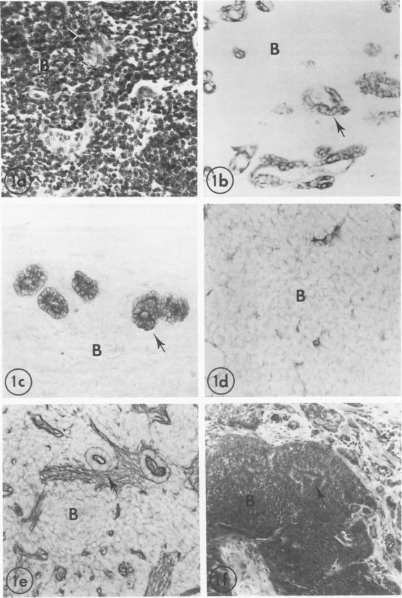
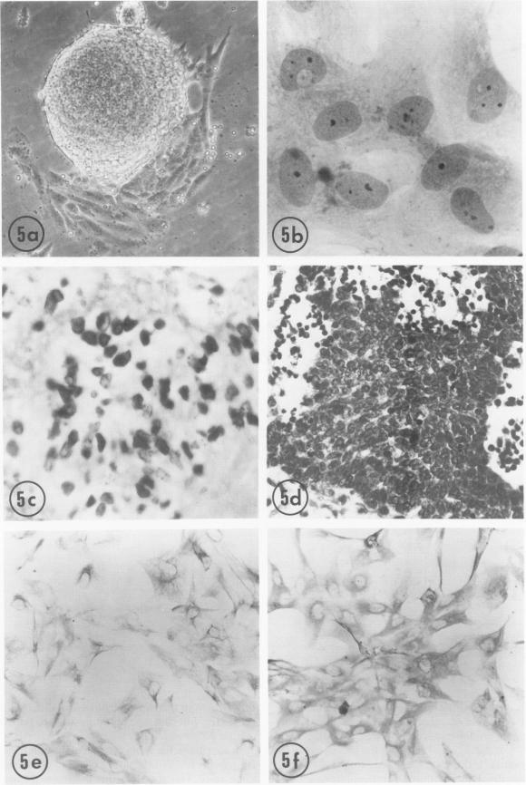
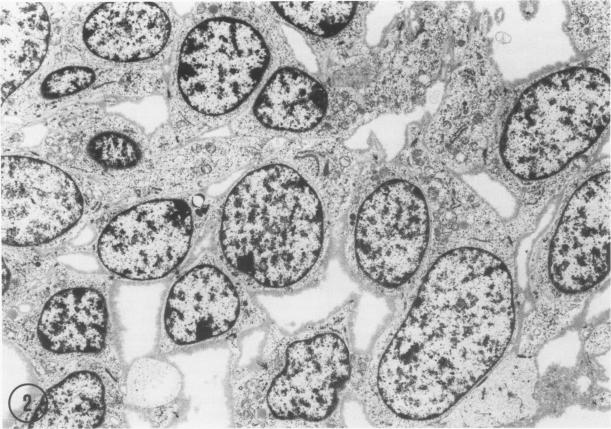
Wilms' tumor has been proposed to originate from a developmental abnormality of the metanephric blastema. This undifferentiated component of Wilms' tumors has previously eluded efforts for in vitro growth. Blastema from a "classical" Wilms' tumor was transplanted into nude mice and passaged through 12 generations of heterotransplantation. Tumors from heterotransplants were grown for 12 serial passages in a serum-free growth medium supplemented with hormones and conditioned media from human kidney proximal tubule cells. The blastema initially grew on a collagen-fetal calf serum matrix as multicellular spheroids, and the cells proliferating from the rim of the spheroids had a flattened shape. Pulse-labeling with bromodeoxyuridine (BrdU) identified the proliferating cell population as blastemal in origin. Except for a loss of extracellular matrix, ultrastructural studies demonstrated morphologic similarities in the cultured cells, compared with the primary tumor and heterotransplants. Lectin histochemical stains for the peanut lectin (PNA) and immunohistochemical stains for cytokeratin (CYTO), vimentin (VIM), and epithelial membrane antigen (EMA) were performed on the original tumor, successive heterotransplants, and cells grown in vitro. The PNA stained the surface of the blastemal cells after sialidase digestion in the original tumor, heterotransplants, and cultured cells. The blastema of the original tumors was negative for CYTO and EMA but reactive for vimentin. This lack of differentiation was maintained in heterotransplants through 12 passages. However, blastemal cells demonstrated coexpression of CYTO and VIM intermediate filaments when grown in a serum-free medium on a matrix material. These studies demonstrate that the blastemal component of Wilms' tumor can be successfully grown in culture, passaged in nude mouse heterotransplants, and shown to undergo early stages of blastemal differentiation in vitro by growth in serum-free medium. This in vitro system provides a model for testing the factors that influence the growth and differentiation of the blastemal component of Wilms' tumors.
肾母细胞瘤被认为起源于后肾胚基的发育异常。肾母细胞瘤的这种未分化成分此前一直未能在体外培养成功。将一个“经典”肾母细胞瘤的胚基移植到裸鼠体内,并经过12代异种移植传代。将异种移植产生的肿瘤在添加了激素和人肾近端小管细胞条件培养基的无血清生长培养基中进行12次连续传代培养。胚基最初在胶原 - 胎牛血清基质上以多细胞球体的形式生长,从球体边缘增殖的细胞呈扁平状。用溴脱氧尿苷(BrdU)进行脉冲标记确定增殖细胞群起源于胚基。除了细胞外基质的丧失外,超微结构研究表明,与原发肿瘤和异种移植相比,培养细胞在形态上具有相似性。对原始肿瘤、连续的异种移植以及体外培养的细胞进行了花生凝集素(PNA)的凝集素组织化学染色和细胞角蛋白(CYTO)、波形蛋白(VIM)及上皮膜抗原(EMA)的免疫组织化学染色。在原始肿瘤、异种移植和培养细胞中,经唾液酸酶消化后,PNA可对胚基细胞表面进行染色。原始肿瘤的胚基对CYTO和EMA呈阴性,但对波形蛋白有反应。这种未分化状态在异种移植中经过12代传代仍得以维持。然而,当在基质材料上的无血清培养基中生长时,胚基细胞显示出CYTO和VIM中间丝的共表达。这些研究表明,肾母细胞瘤的胚基成分能够在培养中成功生长,在裸鼠异种移植中传代,并通过在无血清培养基中生长显示出在体外进行胚基分化的早期阶段。这个体外系统为测试影响肾母细胞瘤胚基成分生长和分化的因素提供了一个模型。