Wuhan Institute for Neuroscience and Neuroengineering, South-Central University for Nationalities, Wuhan, China ; College of Biomedical Engineering, South-Central University for Nationalities, Wuhan, China.
Wuhan Institute for Neuroscience and Neuroengineering, South-Central University for Nationalities, Wuhan, China ; College of Biomedical Engineering, South-Central University for Nationalities, Wuhan, China ; Department of Pharmacology, College of Pharmacy, South-Central University for Nationalities, Wuhan, China.
PLoS One. 2014 Jan 15;9(1):e85643. doi: 10.1371/journal.pone.0085643. eCollection 2014.
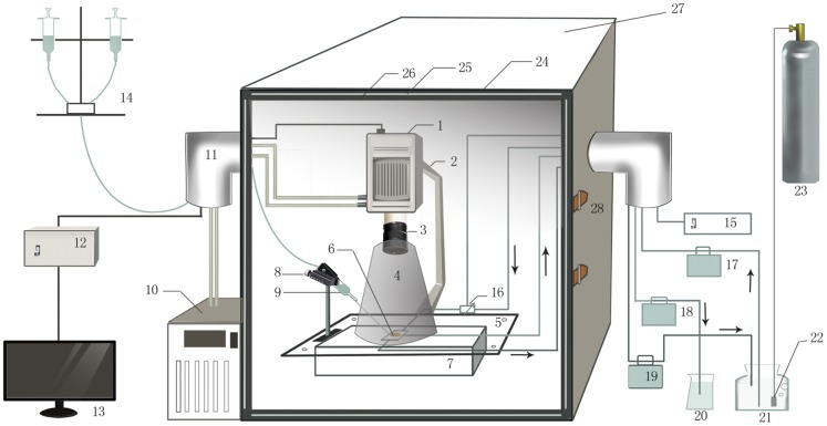
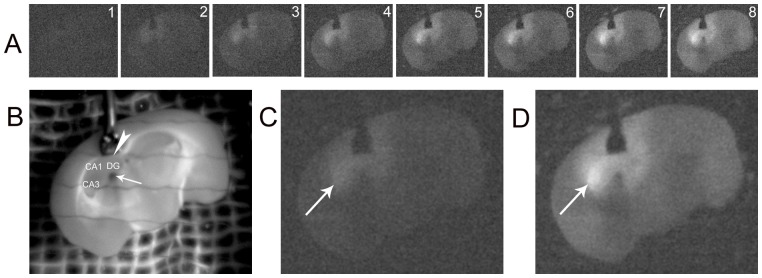
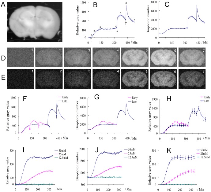
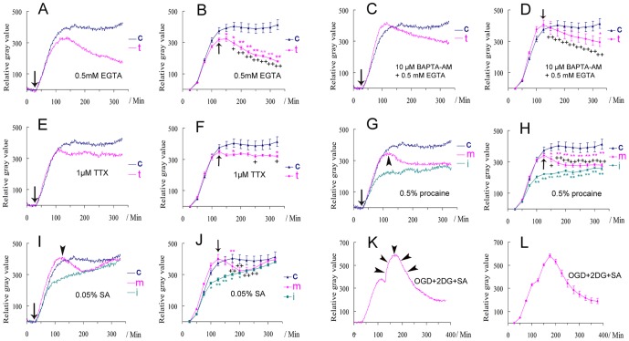
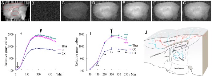
The processing of neural information in neural circuits plays key roles in neural functions. Biophotons, also called ultra-weak photon emissions (UPE), may play potential roles in neural signal transmission, contributing to the understanding of the high functions of nervous system such as vision, learning and memory, cognition and consciousness. However, the experimental analysis of biophotonic activities (emissions) in neural circuits has been hampered due to technical limitations. Here by developing and optimizing an in vitro biophoton imaging method, we characterize the spatiotemporal biophotonic activities and transmission in mouse brain slices. We show that the long-lasting application of glutamate to coronal brain slices produces a gradual and significant increase of biophotonic activities and achieves the maximal effect within approximately 90 min, which then lasts for a relatively long time (>200 min). The initiation and/or maintenance of biophotonic activities by glutamate can be significantly blocked by oxygen and glucose deprivation, together with the application of a cytochrome c oxidase inhibitor (sodium azide), but only partly by an action potential inhibitor (TTX), an anesthetic (procaine), or the removal of intracellular and extracellular Ca(2+). We also show that the detected biophotonic activities in the corpus callosum and thalamus in sagittal brain slices mostly originate from axons or axonal terminals of cortical projection neurons, and that the hyperphosphorylation of microtubule-associated protein tau leads to a significant decrease of biophotonic activities in these two areas. Furthermore, the application of glutamate in the hippocampal dentate gyrus results in increased biophotonic activities in its intrahippocampal projection areas. These results suggest that the glutamate-induced biophotonic activities reflect biophotonic transmission along the axons and in neural circuits, which may be a new mechanism for the processing of neural information.
神经回路中的神经信息处理在神经功能中起着关键作用。生物光子,也称为超弱光发射(UPE),可能在神经信号传递中发挥潜在作用,有助于理解视觉、学习和记忆、认知和意识等神经系统的高级功能。然而,由于技术限制,生物光子活动(发射)在神经回路中的实验分析一直受到阻碍。在这里,我们通过开发和优化体外生物光子成像方法,对小鼠脑片的时空生物光子活动和传输进行了特征描述。我们表明,谷氨酸对冠状脑片的长时间应用会产生生物光子活动的逐渐显著增加,并在大约 90 分钟内达到最大效果,然后持续较长时间(>200 分钟)。谷氨酸引发和/或维持生物光子活动可被氧和葡萄糖剥夺,以及细胞色素 c 氧化酶抑制剂(叠氮化钠)显著阻断,但仅部分被动作电位抑制剂(TTX)、麻醉剂(普鲁卡因)或细胞内和细胞外 Ca(2+)的去除阻断。我们还表明,在矢状脑片中检测到的胼胝体和丘脑中的生物光子活动主要源自皮质投射神经元的轴突或轴突末端,微管相关蛋白 tau 的高度磷酸化会导致这两个区域的生物光子活动显著减少。此外,在海马齿状回中应用谷氨酸会导致其海马内投射区的生物光子活动增加。这些结果表明,谷氨酸诱导的生物光子活动反映了沿着轴突和神经回路的生物光子传输,这可能是神经信息处理的新机制。