Sun Dongchong, Yang Yong, Wei Zhitao, Xu Yong, Zhang Xu, Hong Baofa
Department of Urology, People's Liberation Army General Hospital, Beijing, China.
J Cell Mol Med. 2014 Mar;18(3):434-43. doi: 10.1111/jcmm.12157. Epub 2014 Jan 25.
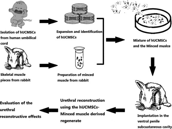
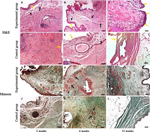
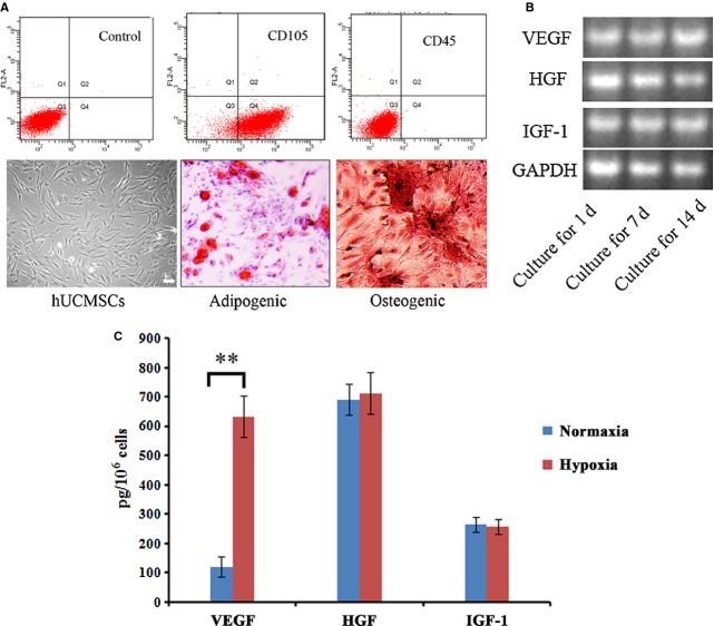
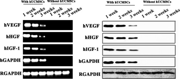
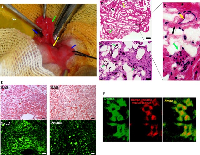
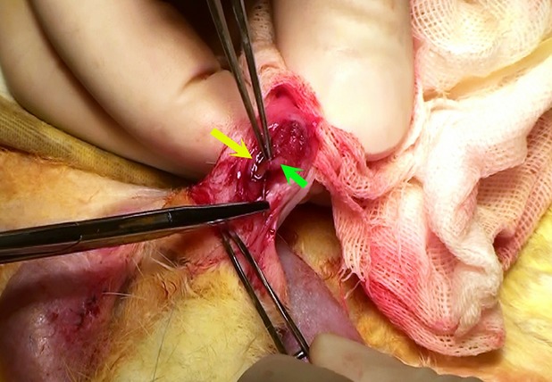
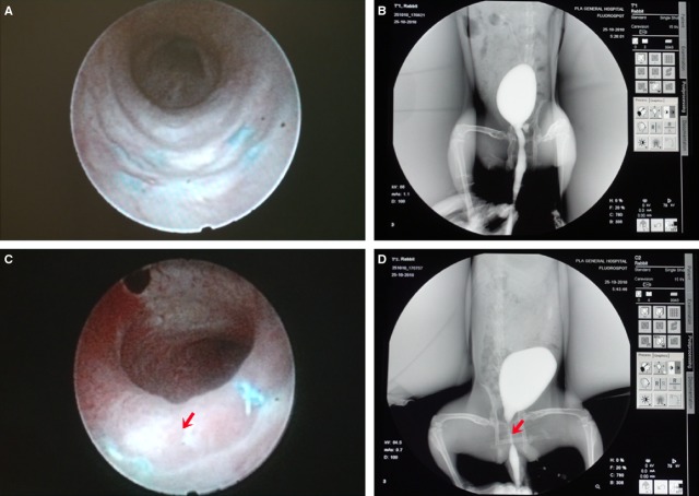
Tissue engineering has brought new hopes for urethral reconstruction. However, the absence of pre-vascularization and the subsequent degradation of materials often lead to the failure of in vivo application. In this study, with the assistance of hypoxia-activated human umbilical cord mesenchymal stem cells (hUCMSCs), pedicled muscle flaps were used as materials and pre-incubated in ventral penile subcutaneous cavity of rabbit for 3 weeks to prepare a pre-vascularized urethral construct. We found that small vessels and muscle fibres were scattered in the construct after 3 weeks' pre-incubation. The construct presented a fibrous reticular structure, which was similar to that of the corpus spongiosum under microscope examination. The produced constructs were then used as a patch graft for reconstruction of the defective rabbit urethra (experimental group), natural muscular patch was used as control (control group). Twelve weeks after the reconstructive surgery, urethrography and urethroscope inspections showed wide calibres of the reconstructed urethra in the experimental group. Histopathological studies revealed that fibrous connective tissues and abundant muscle fibres constituted the main body of the patch-grafted urethra. In contrast, in the control group, only adipose tissue was found in the stenosis-reconstructed urethra, replacing the originally grafted muscular tissue. To our knowledge, this is the first report that successfully constructed a pre-vascularized urethral construct by using hypoxia-activated hUCMSC and pedicled muscle flaps. More importantly, the pre-vascularized construct showed a good performance in urethral reconstruction when applied in vivo. The study provided a novel strategy for tissue engineering of pre-vascularized urethral construct for the defective urethra, representing a further advancement in urethral reconstruction.
组织工程为尿道重建带来了新的希望。然而,缺乏预血管化以及随后材料的降解常常导致体内应用失败。在本研究中,在缺氧激活的人脐带间充质干细胞(hUCMSCs)的辅助下,将带蒂肌瓣用作材料,并在兔阴茎腹侧皮下腔中预孵育3周,以制备预血管化尿道构建体。我们发现,预孵育3周后,构建体中有小血管和肌纤维散在分布。构建体呈现出纤维网状结构,在显微镜检查下与海绵体相似。然后将制备好的构建体用作补片移植材料,用于重建兔尿道缺损(实验组),以天然肌肉补片作为对照(对照组)。重建手术后12周,尿道造影和尿道镜检查显示实验组重建尿道口径宽大。组织病理学研究表明,纤维结缔组织和丰富的肌纤维构成了补片移植尿道的主体。相比之下,在对照组中,狭窄重建的尿道中仅发现脂肪组织,取代了原来移植的肌肉组织。据我们所知,这是首次报道通过使用缺氧激活的hUCMSC和带蒂肌瓣成功构建预血管化尿道构建体。更重要的是,预血管化构建体在体内应用于尿道重建时表现良好。该研究为有缺陷尿道的预血管化尿道构建体的组织工程提供了一种新策略,代表了尿道重建的进一步进展。