Department of Chemistry, University at Albany, State University of New York , 1400 Washington Avenue, Albany, New York 12222, United States.
J Am Chem Soc. 2014 Feb 12;136(6):2302-12. doi: 10.1021/ja407583r. Epub 2014 Jan 31.
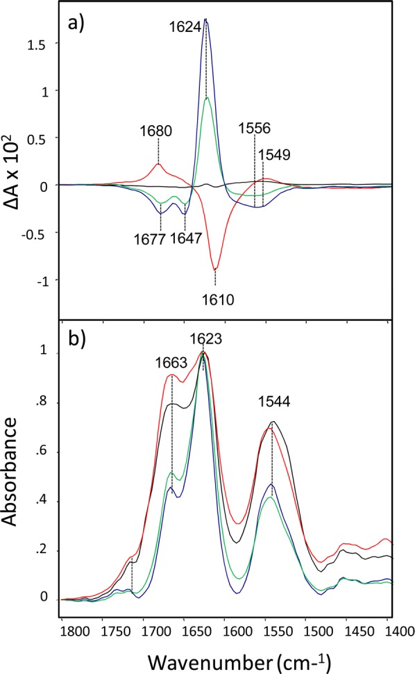
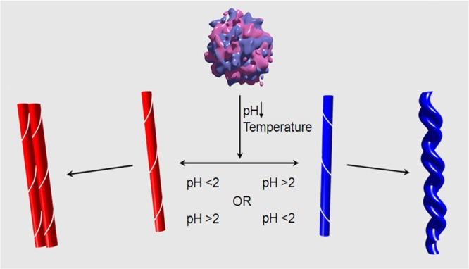
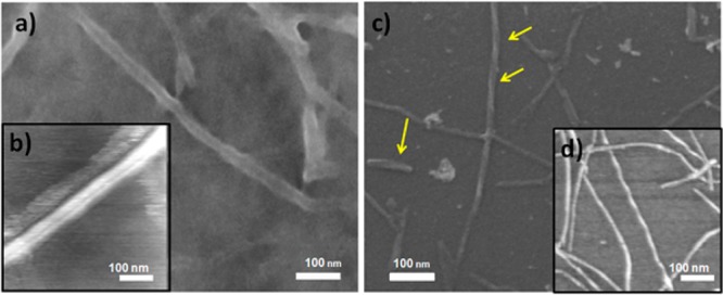
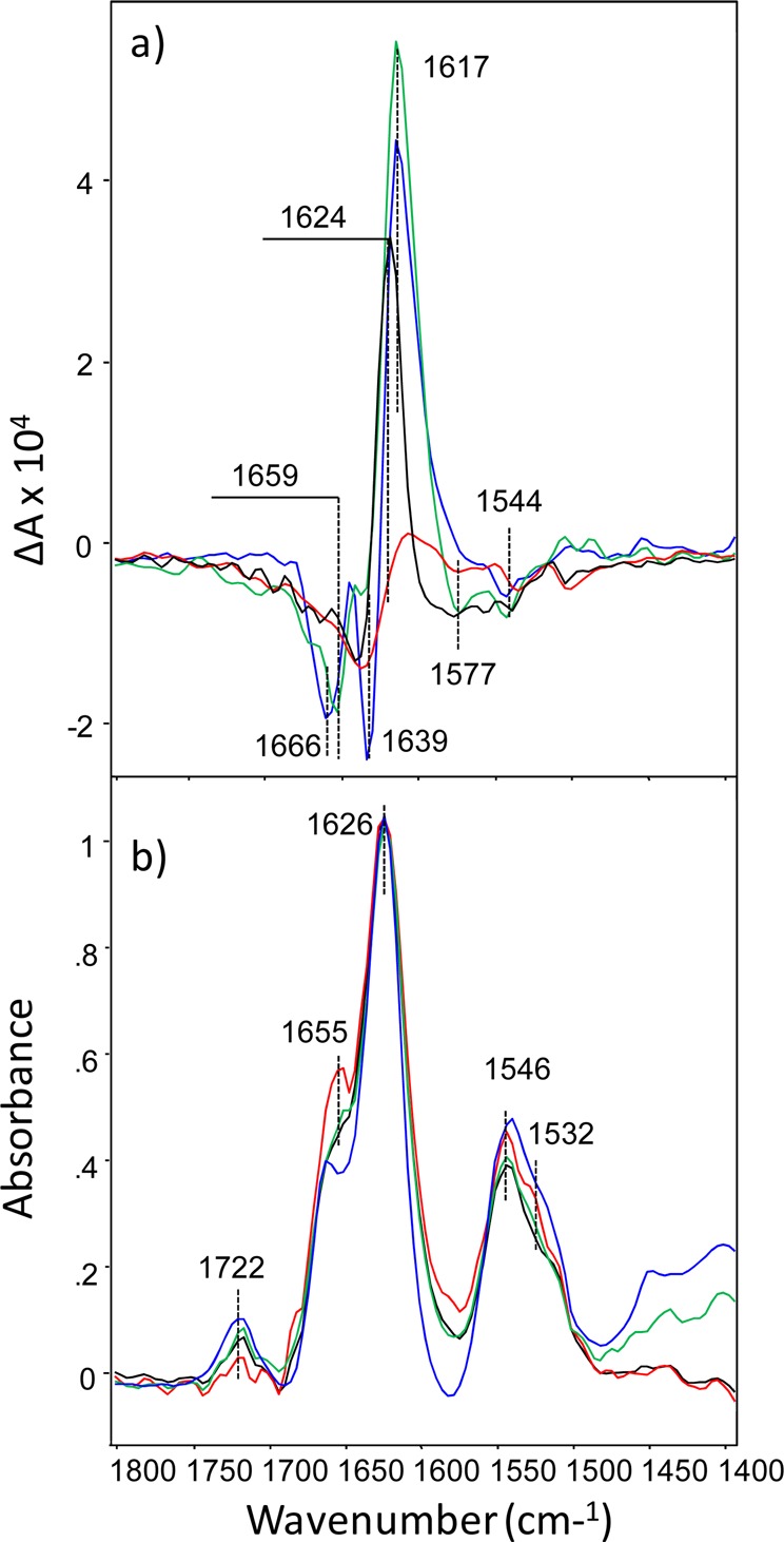
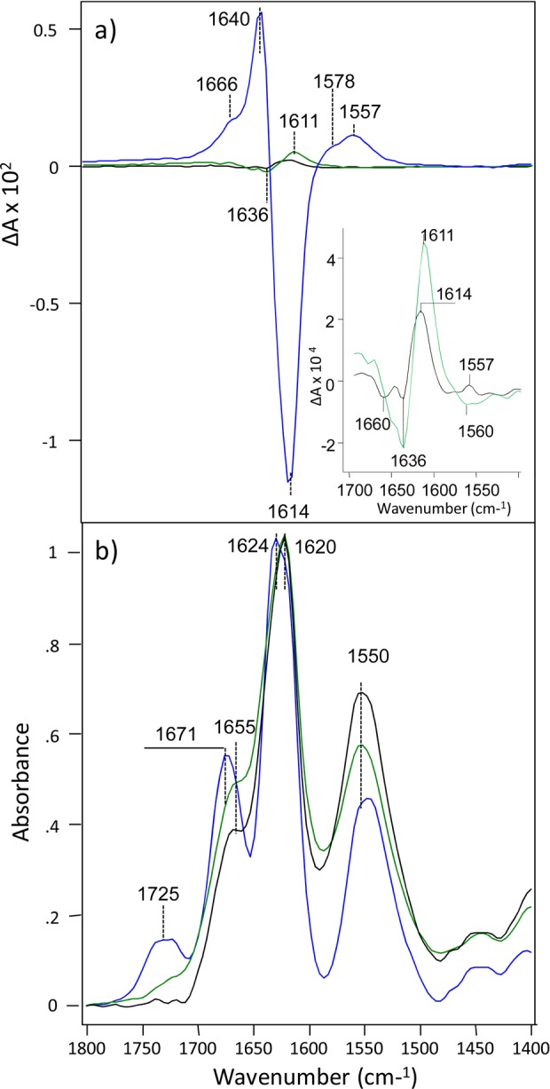
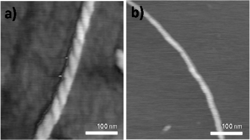
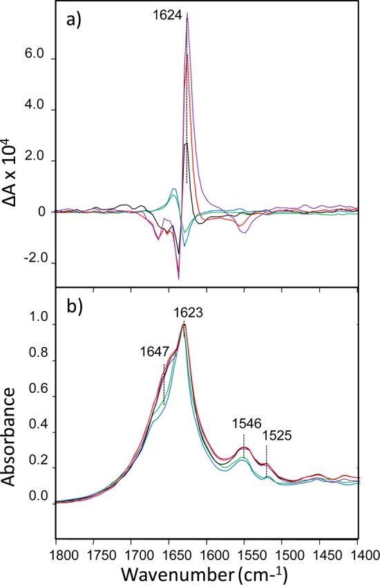
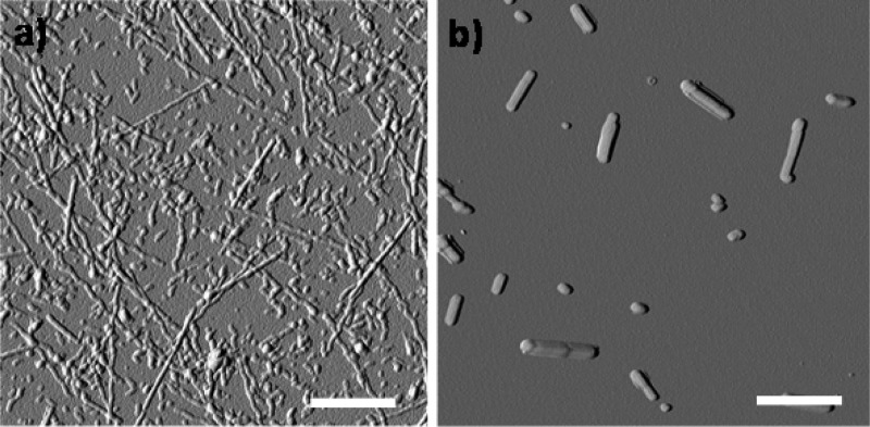
The unique enhanced sensitivity of vibrational circular dichroism (VCD) to the formation and development of amyloid fibrils in solution is extended to four additional fibril-forming proteins or peptides where it is shown that the sign of the fibril VCD pattern correlates with the sense of supramolecular filament chirality and, without exception, to the dominant fibril morphology as observed in AFM or SEM images. Previously for insulin, it has been demonstrated that the sign of the VCD band pattern from filament chirality can be controlled by adjusting the pH of the incubating solution, above pH 2 for "normal" left-hand-helical filaments and below pH 2 for "reversed" right-hand-helical filaments. From AFM or SEM images, left-helical filaments form multifilament braids of left-twisted fibrils while the right-helical filaments form parallel filament rows of fibrils with a flat tape-like morphology, the two major classes of fibril morphology that from deep UV resonance Raman scattering exhibit the same cross-β-core secondary structure. Here we investigate whether fibril supramolecular chirality is the underlying cause of the major morphology differences in all amyloid fibrils by showing that the morphology (twisted versus flat) of fibrils of lysozyme, apo-α-lactalbumin, HET-s (218-289) prion, and a short polypeptide fragment of transthyretin, TTR (105-115), directly correlates to their supramolecular chirality as revealed by VCD. The result is strong evidence that the chiral supramolecular organization of filaments is the principal underlying cause of the morphological heterogeneity of amyloid fibrils. Because fibril morphology is linked to cell toxicity, the chirality of amyloid aggregates should be explored in the widely used in vitro models of amyloid-associated diseases.
振动圆二色性(VCD)对溶液中淀粉样纤维形成和发展的独特增强敏感性扩展到另外四个形成纤维的蛋白质或肽,其中表明纤维 VCD 模式的符号与超分子细丝手性的意义相关,并且无一例外地与在 AFM 或 SEM 图像中观察到的主要纤维形态相关。以前已经证明,对于胰岛素,通过调节孵育溶液的 pH 值,可以控制来自细丝手性的 VCD 带模式的符号,高于 pH 2 时为“正常”左手螺旋细丝,低于 pH 2 时为“反向”右手螺旋细丝。从 AFM 或 SEM 图像中可以看出,左螺旋纤维形成左扭纤维的多纤维辫,而右螺旋纤维形成具有平坦带状形态的平行纤维排,这两种主要的纤维形态,从深紫外共振拉曼散射中表现出相同的交叉-β 核心二级结构。在这里,我们通过表明溶菌酶、apo-α-乳白蛋白、HET-s(218-289)朊病毒和转甲状腺素蛋白 TTR(105-115)短多肽片段的纤维的形态(扭曲与平坦)与其 VCD 揭示的超分子手性直接相关,来研究纤维超分子手性是否是所有淀粉样纤维主要形态差异的根本原因。该结果有力地证明了细丝的手性超分子组织是淀粉样纤维形态异质性的主要原因。由于纤维形态与细胞毒性有关,因此应该在广泛使用的淀粉样相关疾病的体外模型中探索淀粉样聚集物的手性。