• 文献检索
  • 文档翻译
  • 深度研究
  • 学术资讯
  • Suppr Zotero 插件Zotero 插件
  • 邀请有礼
  • 套餐&价格
  • 历史记录
应用&插件
Suppr Zotero 插件Zotero 插件浏览器插件Mac 客户端Windows 客户端微信小程序
定价
高级版会员购买积分包购买API积分包
服务
文献检索文档翻译深度研究API 文档MCP 服务
关于我们
关于 Suppr公司介绍联系我们用户协议隐私条款
关注我们

Suppr 超能文献

核心技术专利:CN118964589B侵权必究
粤ICP备2023148730 号-1Suppr @ 2026

文献检索

告别复杂PubMed语法,用中文像聊天一样搜索,搜遍4000万医学文献。AI智能推荐,让科研检索更轻松。

立即免费搜索

文件翻译

保留排版,准确专业,支持PDF/Word/PPT等文件格式,支持 12+语言互译。

免费翻译文档

深度研究

AI帮你快速写综述,25分钟生成高质量综述,智能提取关键信息,辅助科研写作。

立即免费体验

天然α2-巨球蛋白的结构及其向蛋白酶结合形式的转变。

Structure of native alpha 2-macroglobulin and its transformation to the protease bound form.

作者信息

Bretaudiere J P, Tapon-Bretaudiere J, Stoops J K

机构信息

University of Texas Health Science Center, Department of Pathology and Laboratory Medicine, Houston 77225.

出版信息

Proc Natl Acad Sci U S A. 1988 Mar;85(5):1437-41. doi: 10.1073/pnas.85.5.1437.

DOI:10.1073/pnas.85.5.1437
PMID:2449692
原文链接:https://pmc.ncbi.nlm.nih.gov/articles/PMC279786/
Abstract

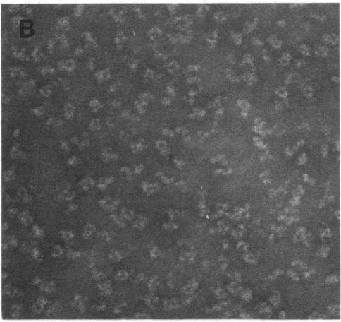
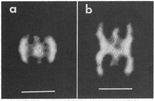
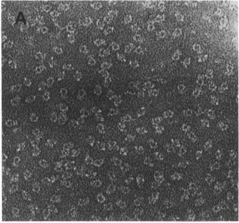
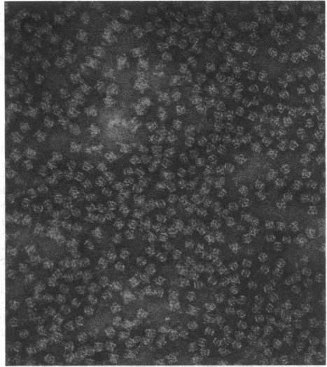
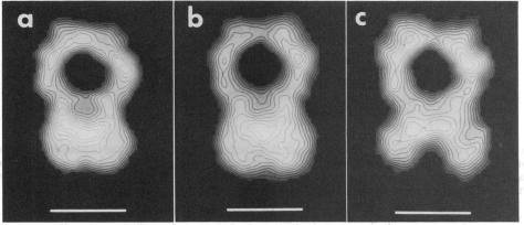
Well-preserved structures of native and alpha-chymotrypsin-bound alpha 2-macroglobulin were obtained by electron microscopy. Computer processing of these images has shown that the native structure has the shape of a padlock 19 nm long. It is proposed that the native alpha 2-macroglobulin consists of the juxtaposition of two protomers with one protomer shaped like a distorted letter "S" and with the other its reverse image, to form a binding site between the two protomers near the bottom of the complex. On cleavage of the subunits with chymotrypsin, the native structure condenses to 16.7 nm and rearranges so that the interaction between the protomers is near the middle. Two images of the alpha 2-macroglobulin-chymotrypsin conjugate were obtained. We suggest that these images represent the end and side view of this complex. Based on the manner in which the native structure is assembled, we propose that the proteolyzed form of alpha 2-macroglobulin is functionally asymmetric in that both protease binding sites reside on the same half of the complex.

摘要

通过电子显微镜获得了天然的以及与α-胰凝乳蛋白酶结合的α2-巨球蛋白的保存完好的结构。对这些图像进行计算机处理后发现,天然结构呈一把长19纳米的挂锁形状。有人提出,天然的α2-巨球蛋白由两个原体并列组成,其中一个原体形状像扭曲的字母“S”,另一个是其镜像,从而在复合物底部附近的两个原体之间形成一个结合位点。用胰凝乳蛋白酶切割亚基后,天然结构收缩至16.7纳米并重新排列,使得原体之间的相互作用靠近中间位置。获得了α2-巨球蛋白-胰凝乳蛋白酶复合物的两张图像。我们认为这些图像代表了该复合物的端面和侧面视图。基于天然结构的组装方式,我们提出α2-巨球蛋白的蛋白水解形式在功能上是不对称的,因为两个蛋白酶结合位点位于复合物的同一半侧。

https://cdn.ncbi.nlm.nih.gov/pmc/blobs/ba4c/279786/6ce06aea8f81/pnas00257-0120-b.jpg
https://cdn.ncbi.nlm.nih.gov/pmc/blobs/ba4c/279786/88a917679cc4/pnas00257-0119-a.jpg
https://cdn.ncbi.nlm.nih.gov/pmc/blobs/ba4c/279786/72941f745adf/pnas00257-0119-b.jpg
https://cdn.ncbi.nlm.nih.gov/pmc/blobs/ba4c/279786/2e8dc3c4294f/pnas00257-0119-c.jpg
https://cdn.ncbi.nlm.nih.gov/pmc/blobs/ba4c/279786/d50f727a6b62/pnas00257-0119-d.jpg
https://cdn.ncbi.nlm.nih.gov/pmc/blobs/ba4c/279786/98b0ec8debea/pnas00257-0120-a.jpg
https://cdn.ncbi.nlm.nih.gov/pmc/blobs/ba4c/279786/6ce06aea8f81/pnas00257-0120-b.jpg

相似文献

1
Structure of native alpha 2-macroglobulin and its transformation to the protease bound form.天然α2-巨球蛋白的结构及其向蛋白酶结合形式的转变。
Proc Natl Acad Sci U S A. 1988 Mar;85(5):1437-41. doi: 10.1073/pnas.85.5.1437.
2
Electron microscope studies of human alpha 2-macroglobulin-chymotrypsin complex: demonstration that the two structures assigned to native and proteolyzed alpha 2-macroglobulin represent two views of the proteolyzed molecule.人α2-巨球蛋白-胰凝乳蛋白酶复合物的电子显微镜研究:证明赋予天然和经蛋白酶解的α2-巨球蛋白的两种结构代表了经蛋白酶解分子的两种视图。
Biochem Biophys Res Commun. 1989 May 30;161(1):216-20. doi: 10.1016/0006-291x(89)91583-0.
3
The three-dimensional structure of the human alpha 2-macroglobulin dimer reveals its structural organization in the tetrameric native and chymotrypsin alpha 2-macroglobulin complexes.人α2-巨球蛋白二聚体的三维结构揭示了其在四聚体天然形式以及胰凝乳蛋白酶-α2-巨球蛋白复合物中的结构组织。
J Biol Chem. 2002 Aug 2;277(31):28031-7. doi: 10.1074/jbc.M202714200. Epub 2002 May 15.
4
Structure-function relationships of human alpha 2-macroglobulin. Three-dimensional structures of native alpha 2-macroglobulin and its methylamine and chymotrypsin derivatives.人α2-巨球蛋白的结构-功能关系。天然α2-巨球蛋白及其甲胺和胰凝乳蛋白酶衍生物的三维结构。
Ann N Y Acad Sci. 1994 Sep 10;737:212-28. doi: 10.1111/j.1749-6632.1994.tb44314.x.
5
Three-dimensional structures of the human alpha 2-macroglobulin-methylamine and chymotrypsin complexes.人α2-巨球蛋白-甲胺和胰凝乳蛋白酶复合物的三维结构。
J Struct Biol. 1992 Nov-Dec;109(3):235-47. doi: 10.1016/1047-8477(92)90036-a.
6
Localization of the two protease binding sites in human alpha 2-macroglobulin.人α2-巨球蛋白中两个蛋白酶结合位点的定位
J Biol Chem. 1981 Jan 25;256(2):547-50.
7
alpha 2-macroglobulin traps a proteinase in the midregion of its arms. An immunoelectron microscopic study.α2-巨球蛋白在其臂的中部捕获蛋白酶。一项免疫电子显微镜研究。
J Biol Chem. 1989 Feb 5;264(4):2350-6.
8
On the structural changes of native human alpha2-macroglobulin upon proteinase entrapment. Three-dimensional structure of the half-transformed molecule.关于蛋白酶捕获后天然人α2-巨球蛋白的结构变化。半转化分子的三维结构。
J Biol Chem. 1998 Apr 10;273(15):8987-93. doi: 10.1074/jbc.273.15.8987.
9
Three-dimensional structure of the human plasmin alpha2-macroglobulin complex.人纤溶酶α2-巨球蛋白复合物的三维结构。
J Struct Biol. 1998 Oct;123(2):124-33. doi: 10.1006/jsbi.1998.4027.
10
Image analysis and three-dimensional model of chymotrypsin-transformed human alpha 2-macroglobulin complexed with a monoclonal antibody specific for this conformation.与针对该构象的单克隆抗体复合的胰凝乳蛋白酶转化的人α2-巨球蛋白的图像分析和三维模型。
Biol Cell. 1988;64(1):45-55. doi: 10.1016/0248-4900(88)90092-5.

引用本文的文献

1
Structural Mechanics of the Alpha-2-Macroglobulin Transformation.α2-巨球蛋白转化的结构力学
J Mol Biol. 2022 Mar 15;434(5):167413. doi: 10.1016/j.jmb.2021.167413. Epub 2021 Dec 20.
2
Inactivation of alpha(2)-Macroglobulin by Activated Human Polymorphonuclear Leukocytes.人活化多形核白细胞对α2-巨球蛋白的灭活作用。
Mediators Inflamm. 1994;3(2):117-23. doi: 10.1155/S0962935194000141.
3
Three-dimensional structure of the Z band in a normal mammalian skeletal muscle.正常哺乳动物骨骼肌中Z带的三维结构。

本文引用的文献

1
Electron microscopy of the conformational changes of alpha 2-macroglobulin from human plasma.人血浆α2-巨球蛋白构象变化的电子显微镜观察。
EMBO J. 1985 Jan;4(1):85-9. doi: 10.1002/j.1460-2075.1985.tb02321.x.
2
Computer averaging of electron micrographs of 40S ribosomal subunits.40S核糖体亚基电子显微照片的计算机平均化处理。
Science. 1981 Dec 18;214(4527):1353-5. doi: 10.1126/science.7313694.
3
Use of multivariate statistics in analysing the images of biological macromolecules.多元统计在生物大分子图像分析中的应用。
J Cell Biol. 1996 May;133(3):571-83. doi: 10.1083/jcb.133.3.571.
4
Structural studies of human alpha 2-macroglobulin: concordance between projected views obtained by negative-stain and cryoelectron microscopy.人α2-巨球蛋白的结构研究:负染和冷冻电子显微镜获得的投影图之间的一致性
J Struct Biol. 1991 Apr;106(2):172-8. doi: 10.1016/1047-8477(91)90086-c.
5
The structure of alpha 2-macroglobulin-methylamine after papain digestion as determined by electron microscopy.经木瓜蛋白酶消化后,通过电子显微镜测定的α2-巨球蛋白-甲胺的结构。
Biochem J. 1990 Sep 1;270(2):291-5. doi: 10.1042/bj2700291.
6
Structure-function relationships of the yeast fatty acid synthase: negative-stain, cryo-electron microscopy, and image analysis studies of the end views of the structure.酵母脂肪酸合酶的结构-功能关系:结构端视图的负染色、冷冻电子显微镜及图像分析研究
Proc Natl Acad Sci U S A. 1992 Jul 15;89(14):6585-9. doi: 10.1073/pnas.89.14.6585.
7
Cloning of the human alpha 2-macroglobulin gene and detection of mutations in two functional domains: the bait region and the thiolester site.人类α2-巨球蛋白基因的克隆及两个功能结构域(诱饵区域和硫酯位点)突变的检测。
Hum Genet. 1992 Jan;88(3):313-9. doi: 10.1007/BF00197266.
Ultramicroscopy. 1981;6(2):187-94. doi: 10.1016/0304-3991(81)90059-0.
4
Primary structure of human alpha 2-macroglobulin. V. The complete structure.人α2-巨球蛋白的一级结构。V. 完整结构。
J Biol Chem. 1984 Jul 10;259(13):8318-27.
5
Structure of alpha 2-macroglobulin in solution and its interaction with proteases: an X-ray scattering study using the contrast variation method.溶液中α2-巨球蛋白的结构及其与蛋白酶的相互作用:使用对比变化法的X射线散射研究。
Ann N Y Acad Sci. 1983;421:98-111. doi: 10.1111/j.1749-6632.1983.tb18096.x.
6
Reaction of human alpha 2-macroglobulin half-molecules with plasmin as a probe of protease binding site structure.人α2-巨球蛋白半分子与纤溶酶的反应:作为蛋白酶结合位点结构的探针
Biochemistry. 1983 Oct 11;22(21):4933-40. doi: 10.1021/bi00290a009.
7
Interaction of alpha 2-macroglobulin with trypsin, chymotrypsin, plasmin, and papain.α2-巨球蛋白与胰蛋白酶、胰凝乳蛋白酶、纤溶酶和木瓜蛋白酶的相互作用。
Arch Biochem Biophys. 1983 Feb 15;221(1):261-70. doi: 10.1016/0003-9861(83)90143-1.
8
Multivariate statistical analysis of ribosome electron micrographs. L and R lateral views of the 40 S subunit from HeLa cells.
J Mol Biol. 1982 Oct 15;161(1):107-33. doi: 10.1016/0022-2836(82)90281-9.
9
Physical properties of human alpha 2-macroglobulin following reaction with methylamine and trypsin.人α2-巨球蛋白与甲胺和胰蛋白酶反应后的物理性质
Biochim Biophys Acta. 1982 Aug 10;705(3):306-14. doi: 10.1016/0167-4838(82)90252-7.
10
Computer averaging of single molecules of alpha 2-macroglobulin and the alpha 2-macroglobulin/trypsin complex.α2-巨球蛋白单分子以及α2-巨球蛋白/胰蛋白酶复合物的计算机平均化处理
Hoppe Seylers Z Physiol Chem. 1982 Aug;363(8):803-12. doi: 10.1515/bchm2.1982.363.2.803.