Department of Radiology, Yonsei University College of Medicine, Gangnam Severance Hospital, 211 Eonju-ro, Gangnam-gu, Seoul 135-720, Korea.
Yonsei Med J. 2014 Mar;55(2):339-44. doi: 10.3349/ymj.2014.55.2.339.
To analyze which sonographic features of thyroid nodules with macrocalcifications were predictable of thyroid malignancy.
We reviewed sonographic findings of 854 macrocalcified thyroid nodules in patients who underwent fine needle aspiration biopsy between December 2009 and January 2011. There were 171 non-diagnostic aspirations, 34 nodules with category 3, 4, 5 based on Bethesda system, which were not confirmed by surgery, and these nodules were excluded from the analysis. Sonographic characteristics of the macrocalcifications including its thickness, interruption, and existence of soft tissue rim outside the macrocalcification were analyzed. Other sonographic characteristics of nodules such as shape, margin, composition, echo pattern, vascularity, and underlying parenchymal echogenicity were also evaluated. The correlation of sonographic features with cytopathologic results and the diagnostic performance of sonographic features for the prediction of malignancy were analyzed.
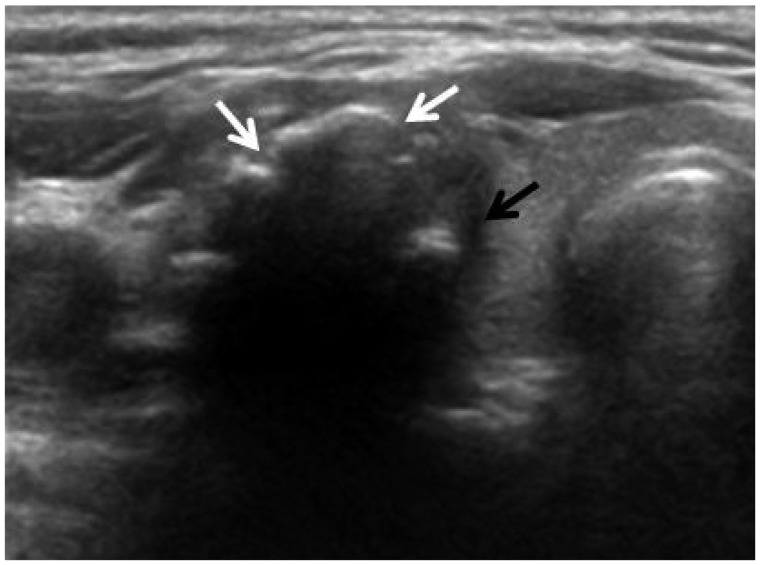
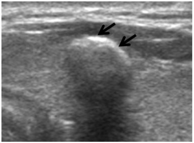
Among 649 nodules, 179 (27.6%) nodules were malignant and 470 (72.4%) nodules were benign. Among the features of the macrocalcification, interruption, irregular thickness, or the presence of soft tissue outside calcification rim were associated with malignancy (p<0.001). A high sensitivity and negative predictive values for the prediction of malignancy was found in sonographic characteristics of irregular thickness (92.2% and 91.0%, respectively) and the presence of soft tissue (88.5% and 88.8%, respectively).
Sonographic characteristics of macrocalcification such as interruption, irregular thickness and the presence of soft tissue rim were associated with malignancy in thyroid nodules with macrocalcifications.
分析伴有大钙化的甲状腺结节的超声特征哪些可预测甲状腺恶性肿瘤。
我们回顾了 2009 年 12 月至 2011 年 1 月期间接受细针抽吸活检的 854 个大钙化甲状腺结节患者的超声检查结果。其中 171 例为非诊断性抽吸,34 例为贝塞斯达系统分类为 3、4、5 类的结节,但未通过手术证实,这些结节被排除在分析之外。分析了大钙化的超声特征,包括其厚度、中断和大钙化外是否存在软组织边缘。还评估了结节的其他超声特征,如形状、边界、成分、回声模式、血流和基础实质回声。分析了超声特征与细胞学结果的相关性,以及超声特征对恶性肿瘤预测的诊断性能。
在 649 个结节中,179 个(27.6%)结节为恶性,470 个(72.4%)结节为良性。在大钙化的特征中,中断、不规则厚度或钙化边缘外存在软组织与恶性肿瘤有关(p<0.001)。不规则厚度(92.2%和 91.0%)和存在软组织(88.5%和 88.8%)的超声特征对恶性肿瘤的预测具有较高的敏感性和阴性预测值。
伴有大钙化的甲状腺结节中,大钙化的超声特征如中断、不规则厚度和存在软组织边缘与恶性肿瘤有关。