Department of Ophthalmology/Kresge Eye Institute, Wayne State University School of Medicine, 4717 St, Antoine, Detroit, MI 48201, USA.
J Neuroinflammation. 2014 Feb 18;11:33. doi: 10.1186/1742-2094-11-33.
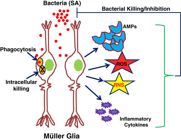
We have previously shown that, in response to microbial infection, activated Müller glia secrete inflammatory cytokines/chemokines and exhibit antimicrobial properties. The aim of this study is to understand the mechanisms and the key components involved in this response.
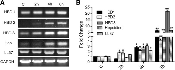
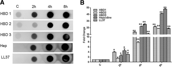
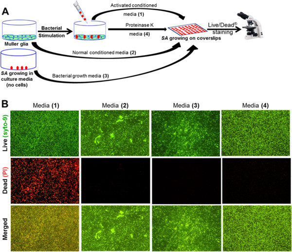
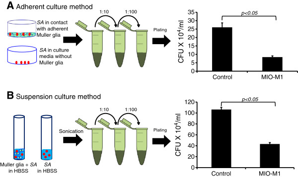
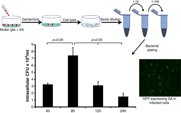
Immortalized human retinal Müller glia (MIO-M1 cells) were challenged with Staphylococcus (S) aureus, the leading cause of severe intraocular infection followed by RT2 profile PCR array analysis. The expression of human β-defensin 1 (HBD1), 2 (HBD2), 3 (HBD3), hepcidine and cathelicidin LL37 was checked by RT-PCR and quantified by Taqman qPCR. The expression of AMPs was confirmed at protein level by dot-blot analysis. The production of ROS was measured by dicholoro-dihydro-fluorescein diacetate (DCFH-DA) staining by flow cytometry as well as fluorescence microscopy. The level of nitric oxide (NO) was measured by measuring a stable metabolite, nitrite using the Griess reagent. In vitro killing assay was performed by Live/Dead BacLight staining as well as by dilution plating in suspension and adherent conditions following S. aureus infection. Phagocytosis was measured by CFU enumeration following infection.
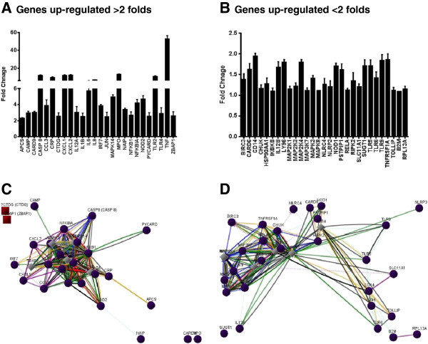
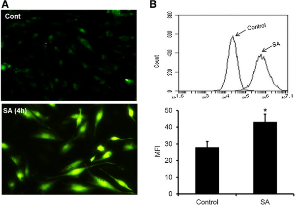
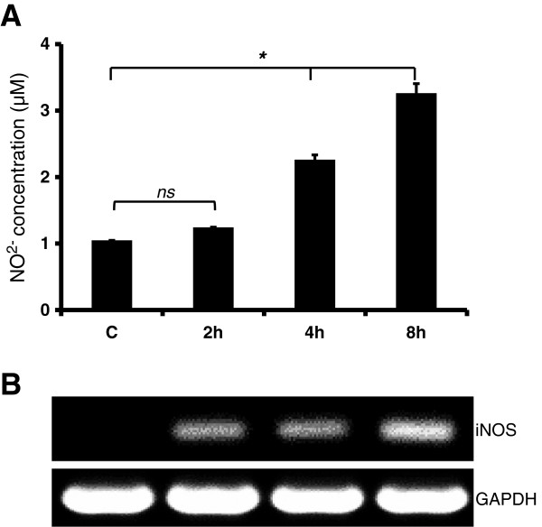
PCR array data showed that, in comparison to uninfected control cells, bacterial challenge significantly (> two-fold) induced the expression of 26 genes involved in cytokine/chemokine, antimicrobials, Toll-like receptor, apoptotic, and NF-κB signaling. RT-PCR analysis showed time-dependent increased expression of HBD1, HBD2, HBD3, LL-37, and hepcidin mRNA in bacteria-challenged Müller glia. The expression of these antimicrobial molecules was also increased at the protein level in the culture supernatant, as detected by dot-blot analysis. Additionally, the bacteria-stimulated Müller glia were found to produce reactive oxygen (ROS) and reactive nitrogen (RNS) species. In vitro, killing assays revealed that Müller glia exhibited bactericidal activity against S. aureus in both adherent and suspension cultures. Furthermore, our data demonstrated that Müller glia can phagocytize and kill the bacteria in a time-dependent manner.
These data suggest that retinal Müller glia behave like classical innate immune cells by producing a variety of antimicrobial molecules in response to bacterial challenge, suggesting their pivotal role in retinal innate defense.
我们之前已经表明,在受到微生物感染后,激活的 Müller 胶质细胞会分泌炎症细胞因子/趋化因子,并表现出抗菌特性。本研究的目的是了解这种反应所涉及的机制和关键成分。
用金黄色葡萄球菌(S. aureus)刺激永生化人视网膜 Müller 胶质细胞(MIO-M1 细胞),然后进行 RT2 谱 PCR 阵列分析。通过 RT-PCR 检查人β防御素 1(HBD1)、2(HBD2)、3(HBD3)、hepcidin 和 cathelicidin LL37 的表达,并通过 Taqman qPCR 进行定量。通过斑点印迹分析确认 AMPs 在蛋白质水平上的表达。通过流式细胞术和荧光显微镜用二氯二氢荧光素二乙酸酯(DCFH-DA)染色测量活性氧(ROS)的产生。通过测量使用 Griess 试剂的稳定代谢物亚硝酸盐来测量一氧化氮(NO)的水平。在金黄色葡萄球菌感染后进行活/死 BacLight 染色以及悬浮和贴壁条件下的稀释平板进行体外杀伤测定。通过感染后的 CFU 计数测量吞噬作用。
PCR 阵列数据显示,与未感染对照细胞相比,细菌刺激显著(>两倍)诱导了 26 个参与细胞因子/趋化因子、抗菌剂、Toll 样受体、凋亡和 NF-κB 信号的基因的表达。RT-PCR 分析显示,在细菌刺激的 Müller 胶质细胞中,HBD1、HBD2、HBD3、LL-37 和 hepcidin mRNA 的表达呈时间依赖性增加。通过斑点印迹分析,还在培养上清液中检测到这些抗菌分子的蛋白质水平增加。此外,发现细菌刺激的 Müller 胶质细胞产生活性氧(ROS)和活性氮(RNS)物质。在体外,杀伤测定表明 Müller 胶质细胞在贴壁和悬浮培养中均对金黄色葡萄球菌具有杀菌活性。此外,我们的数据表明 Müller 胶质细胞可以以时间依赖性方式吞噬和杀死细菌。
这些数据表明,视网膜 Müller 胶质细胞通过在受到细菌刺激时产生多种抗菌分子来表现出典型的先天免疫细胞特性,表明它们在视网膜先天防御中具有关键作用。