Watt F M, Jordan P W, O'Neill C H
Keratinocyte Laboratory, Imperial Cancer Research Fund, London, United Kingdom.
Proc Natl Acad Sci U S A. 1988 Aug;85(15):5576-80. doi: 10.1073/pnas.85.15.5576.
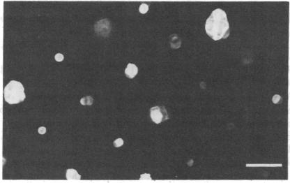
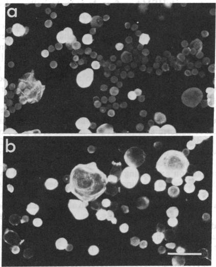
Cultures of human epidermal keratinocytes provide a useful experimental model with which to study the factors that regulate cell proliferation and terminal differentiation. One situation that is known to trigger premature terminal differentiation is suspension culture, when keratinocytes are deprived of substratum and intercellular contact. We have now investigated whether area of substratum contact, and hence cell shape, can regulate terminal differentiation. Keratinocytes were grown on circular adhesive islands that prevented cell-cell contact. By varying island area we could vary cell shape from fully spread to almost spherical. We found that when substratum contact was restricted, DNA synthesis was inhibited and expression of involucrin, a marker of terminal differentiation, was stimulated. Inhibition of proliferation was not a sufficient stimulus for involucrin synthesis in fully spread cells. When DNA synthesis and involucrin expression were plotted against contact area, classic dose-response curves were obtained. Thus cell shape acts as a signal for the terminal differentiation of keratinocytes in culture.
人表皮角质形成细胞培养提供了一个有用的实验模型,可用于研究调节细胞增殖和终末分化的因素。已知一种能触发过早终末分化的情况是悬浮培养,即角质形成细胞被剥夺基质和细胞间接触时。我们现在研究了基质接触面积,进而细胞形状,是否能调节终末分化。角质形成细胞在圆形黏附岛上生长,这些岛可防止细胞间接触。通过改变岛的面积,我们可以使细胞形状从完全铺展变为几乎球形。我们发现,当基质接触受到限制时,DNA合成受到抑制,而终末分化标志物兜甲蛋白的表达受到刺激。在完全铺展的细胞中,增殖抑制并非兜甲蛋白合成的充分刺激因素。当将DNA合成和兜甲蛋白表达与接触面积作图时,得到了典型的剂量反应曲线。因此,细胞形状在培养中作为角质形成细胞终末分化的信号。