Babot Marion, Birch Amanda, Labarbuta Paola, Galkin Alexander
Queen's University Belfast, School of Biological Sciences, Medical Biology Centre, 97 Lisburn Road, Belfast BT9 7BL, UK.
Queen's University Belfast, School of Biological Sciences, Medical Biology Centre, 97 Lisburn Road, Belfast BT9 7BL, UK.
Biochim Biophys Acta. 2014 Jul;1837(7):1083-92. doi: 10.1016/j.bbabio.2014.02.018. Epub 2014 Feb 22.
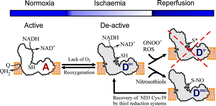
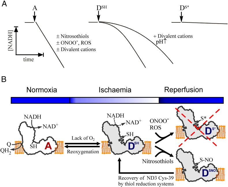
Oxidation of NADH in the mitochondrial matrix of aerobic cells is catalysed by mitochondrial complex I. The regulation of this mitochondrial enzyme is not completely understood. An interesting characteristic of complex I from some organisms is the ability to adopt two distinct states: the so-called catalytically active (A) and the de-active, dormant state (D). The A-form in situ can undergo de-activation when the activity of the respiratory chain is limited (i.e. in the absence of oxygen). The mechanisms and driving force behind the A/D transition of the enzyme are currently unknown, but several subunits are most likely involved in the conformational rearrangements: the accessory subunit 39kDa (NDUFA9) and the mitochondrially encoded subunits, ND3 and ND1. These three subunits are located in the region of the quinone binding site. The A/D transition could represent an intrinsic mechanism which provides a fast response of the mitochondrial respiratory chain to oxygen deprivation. The physiological role of the accumulation of the D-form in anoxia is most probably to protect mitochondria from ROS generation due to the rapid burst of respiration following reoxygenation. The de-activation rate varies in different tissues and can be modulated by the temperature, the presence of free fatty acids and divalent cations, the NAD(+)/NADH ratio in the matrix, the presence of nitric oxide and oxygen availability. Cysteine-39 of the ND3 subunit, exposed in the D-form, is susceptible to covalent modification by nitrosothiols, ROS and RNS. The D-form in situ could react with natural effectors in mitochondria or with pharmacological agents. Therefore the modulation of the re-activation rate of complex I could be a way to ameliorate the ischaemia/reperfusion damage. This article is part of a Special Issue entitled: 18th European Bioenergetic Conference. Guest Editors: Manuela Pereira and Miguel Teixeira.
有氧细胞线粒体基质中NADH的氧化由线粒体复合物I催化。这种线粒体酶的调节机制尚未完全明确。某些生物体中复合物I的一个有趣特性是能够呈现两种不同状态:即所谓的催化活性态(A)和失活的休眠态(D)。当呼吸链活性受限(即缺氧时),原位的A态可发生失活。目前尚不清楚该酶A/D转变背后的机制和驱动力,但可能有几个亚基参与了构象重排:辅助亚基39kDa(NDUFA9)以及线粒体编码的亚基ND3和ND1。这三个亚基位于醌结合位点区域。A/D转变可能代表一种内在机制,可使线粒体呼吸链对缺氧做出快速反应。缺氧时D态积累的生理作用很可能是保护线粒体免受复氧后呼吸快速爆发所产生的活性氧的损伤。失活速率在不同组织中有所不同,并且可受到温度、游离脂肪酸和二价阳离子的存在、基质中NAD(+)/NADH比值、一氧化氮的存在以及氧供应情况的调节。ND3亚基的半胱氨酸-39在D态时暴露在外,易受到亚硝基硫醇、活性氧和活性氮的共价修饰。原位的D态可能会与线粒体中的天然效应物或药物发生反应。因此,调节复合物I的再激活速率可能是减轻缺血/再灌注损伤的一种方法。本文是名为:第18届欧洲生物能量学会议的特刊的一部分。客座编辑:曼努埃拉·佩雷拉和米格尔·特谢拉。