Arai Toshio, Akao Nobuaki, Seki Takenori, Kumagai Takashi, Ishikawa Hirofumi, Ohta Nobuo, Hirata Nobuto, Nakaji So, Yamauchi Kenji, Hirai Mitsuru, Shiratori Toshiyasu, Kobayashi Masayoshi, Fujii Hiroyuki, Ishii Eiji, Naito Mikio, Saitoh Shin-ichi, Yamaguchi Toshikazu, Shibata Nobumitsu, Shimo Masamune, Tokiwa Toshihiro
Department of Environmental Parasitology, Graduate School of Tokyo Medical and Dental University, Bunkyo-ku, Tokyo, Japan ; Department of Gastroenterology, Toukatsu Hospital, Nagareyama-shi, Chiba, Japan.
Department of Environmental Parasitology, Graduate School of Tokyo Medical and Dental University, Bunkyo-ku, Tokyo, Japan.
PLoS One. 2014 Feb 28;9(2):e89188. doi: 10.1371/journal.pone.0089188. eCollection 2014.
Anisakiasis is a parasitic disease caused primarily by Anisakis spp. larvae in Asia and in Western countries. The aim of this study was to investigate the genotype of Anisakis larvae endoscopically removed from Middle Eastern Japanese patients and to determine whether mucosal atrophy affects the risk of penetration in gastric anisakiasis.
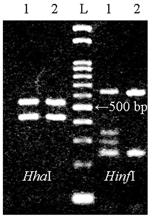
In this study, 57 larvae collected from 44 patients with anisakiasis (42 gastric and 2 colonic anisakiasis) were analyzed retrospectively. Genotyping was confirmed by restriction fragment length polymorphism (RFLP) analysis of ITS regions and by sequencing the mitochondrial small subunit (SSU) region. In the cases of gastric anisakiasis, correlation analyses were conducted between the frequency of larval penetration in normal/atrophic area and the manifestation of clinical symptoms.
Nearly all larvae were A. simplex seusu stricto (s.s.) (99%), and one larva displayed a hybrid genotype. The A. simplex larvae penetrated normal mucosa more frequently than atrophic area (p = 0.005). Finally, patients with normal mucosa infection were more likely to exhibit clinical symptoms than those with atrophic mucosa infection (odds ratio, 6.96; 95% confidence interval, 1.52-31.8).
In Japan, A. simplex s.s. is the main etiological agent of human anisakiasis and tends to penetrate normal gastric mucosa. Careful endoscopic examination of normal gastric mucosa, particularly in the greater curvature of the stomach will improve the detection of Anisakis larvae.
异尖线虫病是一种主要由异尖线虫属幼虫引起的寄生虫病,在亚洲和西方国家均有发生。本研究旨在调查从中东日本患者内镜下取出的异尖线虫幼虫的基因型,并确定黏膜萎缩是否会影响胃异尖线虫病的侵入风险。
本研究对从44例异尖线虫病患者(42例胃异尖线虫病和2例结肠异尖线虫病)收集的57条幼虫进行回顾性分析。通过对ITS区域的限制性片段长度多态性(RFLP)分析和线粒体小亚基(SSU)区域测序来确认基因分型。对于胃异尖线虫病病例,对正常/萎缩区域幼虫侵入频率与临床症状表现之间进行相关性分析。
几乎所有幼虫均为狭义简单异尖线虫(99%),1条幼虫表现出杂交基因型。简单异尖线虫幼虫侵入正常黏膜的频率高于萎缩区域(p = 0.005)。最后,正常黏膜感染患者比萎缩黏膜感染患者更易出现临床症状(优势比,6.96;95%置信区间,1.52 - 31.8)。
在日本,狭义简单异尖线虫是人类异尖线虫病的主要病原体,且倾向于侵入正常胃黏膜。对正常胃黏膜进行仔细的内镜检查,尤其是在胃大弯处,将提高异尖线虫幼虫的检出率。