Wang Lei, Ye Tingjun, Deng Lianfu, Shao Jin, Qi Jin, Zhou Qi, Wei Li, Qiu Shijing
Shanghai Key Laboratory for Prevention and Treatment of Bone and Joint Diseases with Integrated Chinese-Western Medicine, Department of Orthopedics, Shanghai Institute of Traumatology and Orthopedics, Ruijin Hospital, Shanghai Jiaotong University School of Medicine, Shanghai, PR China.
Shanghai Key Laboratory for Prevention and Treatment of Bone and Joint Diseases with Integrated Chinese-Western Medicine, Department of Orthopedics, Shanghai Institute of Traumatology and Orthopedics, Ruijin Hospital, Shanghai Jiaotong University School of Medicine, Shanghai, PR China ; Bone and Mineral Research Laboratory, Henry Ford Hospital, Detroit, Michigan, United States of America.
PLoS One. 2014 Feb 20;9(2):e89343. doi: 10.1371/journal.pone.0089343. eCollection 2014.
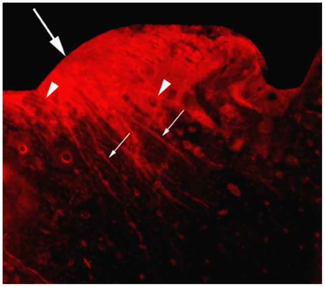
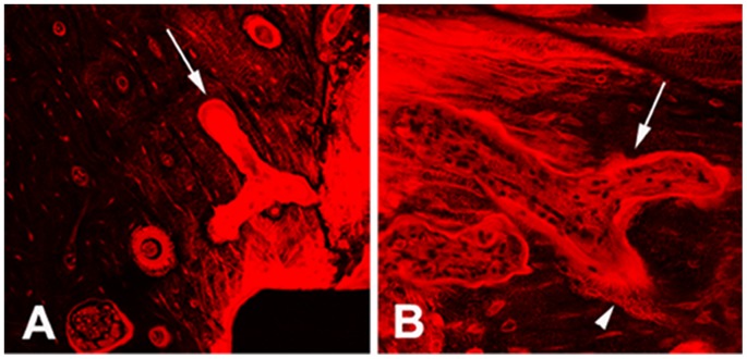
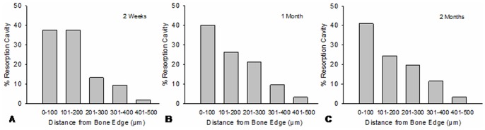
Up to date, little is known about the repair mode of microdamage in osteonal cortical bone resulting from bone screw implantation. In this study, self-tapping titanium cortical bone screws were inserted into the tibial diaphyses of 24 adult male rabbits. The animals were sacrificed at 1 day, 2 weeks, 1 month and 2 months after surgery. Histomorphometric measurement and confocal microscopy were performed on basic fuchsin stained bone sections to examine the morphological characteristics of microdamage, bone resorption activity and spatial relationship between microdamage and bone resorption. Diffuse and linear cracks were coexisted in peri-screw bone. Intracortical bone resorption was significantly increased 2 weeks after screw installation and reach to the maximum at 1 month. There was no significant difference in bone resorption between 1-month and 2-months groups. Microdamage was significantly decreased within 1 month after surgery. Bone resorption was predisposed to occur in the region of <100 µm from the bone-screw interface, where had extensive diffuse damage mixed with linear cracks. Different patterns of resorption cavities appeared in peri-screw bone. These data suggest that 1) the complex microdamage composed of diffuse damage and linear cracks is a strong stimulator for initiating targeted bone remodeling; 2) bone resorption activities taking place on the surfaces of differently oriented Haversian and Volkmann canals work in a team for the repair of extensive microdamage; 3) targeted bone remodeling is a short-term reaction to microdamage and thereby it may not be able to remove all microdamage resulting from bone screw insertion.
到目前为止,关于骨螺钉植入导致的骨单位皮质骨微损伤的修复模式知之甚少。在本研究中,将自攻式钛皮质骨螺钉插入24只成年雄性兔的胫骨干。在手术后1天、2周、1个月和2个月处死动物。对碱性品红染色的骨切片进行组织形态计量学测量和共聚焦显微镜检查,以研究微损伤的形态特征、骨吸收活性以及微损伤与骨吸收之间的空间关系。螺钉周围骨中同时存在弥漫性和线性裂纹。螺钉植入后2周,皮质内骨吸收显著增加,并在1个月时达到最大值。1个月和2个月组之间的骨吸收没有显著差异。手术后1个月内微损伤显著减少。骨吸收易发生在距骨螺钉界面<100 µm的区域,该区域有广泛的弥漫性损伤并伴有线性裂纹。螺钉周围骨中出现了不同模式的吸收腔。这些数据表明:1)由弥漫性损伤和线性裂纹组成的复杂微损伤是启动靶向骨重塑的强烈刺激因素;2)在不同方向的哈弗斯管和福尔克曼管表面发生的骨吸收活动协同作用以修复广泛的微损伤;3)靶向骨重塑是对微损伤的短期反应,因此它可能无法消除骨螺钉插入导致的所有微损伤。