Wang Bo, Kagemann Larry, Schuman Joel S, Ishikawa Hiroshi, Bilonick Richard A, Ling Yun, Sigal Ian A, Nadler Zach, Francis Andrew, Sandrian Michelle G, Wollstein Gadi
UPMC Eye Center, Eye and Ear Institute, Ophthalmology and Visual Science Research Center, Department of Ophthalmology, University of Pittsburgh School of Medicine, Pittsburgh, Pennsylvania, United States of America; Department of Bioengineering, Swanson School of Engineering, University of Pittsburgh, Pittsburgh, Pennsylvania, United States of America; Department of Biostatistics, Graduate School of Public Health, University of Pittsburgh, Pittsburgh, Pennsylvania, United States of America.
UPMC Eye Center, Eye and Ear Institute, Ophthalmology and Visual Science Research Center, Department of Ophthalmology, University of Pittsburgh School of Medicine, Pittsburgh, Pennsylvania, United States of America; Department of Bioengineering, Swanson School of Engineering, University of Pittsburgh, Pittsburgh, Pennsylvania, United States of America.
PLoS One. 2014 Mar 3;9(3):e90690. doi: 10.1371/journal.pone.0090690. eCollection 2014.
To investigate gold nanorods (GNRs) as a contrast agent to enhance Doppler optical coherence tomography (OCT) imaging of the intrascleral aqueous humor outflow.
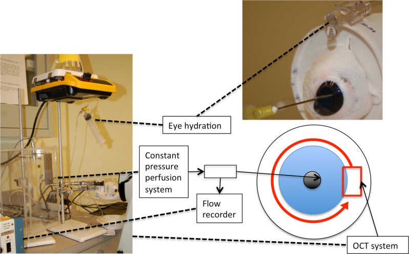
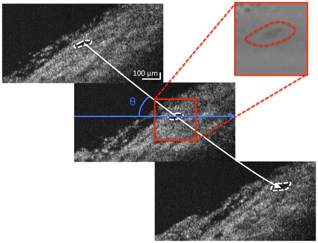
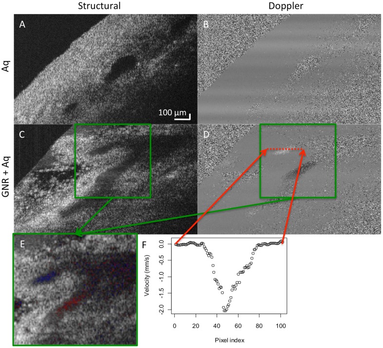
A serial dilution of GNRs was scanned with a spectral-domain OCT device (Bioptigen, Durham, NC) to visualize Doppler signal. Doppler measurements using GNRs were validated using a controlled flow system. To demonstrate an application of GNR enhanced Doppler, porcine eyes were perfused at constant pressure with mock aqueous alone or 1.0×10(12) GNR/mL mixed with mock aqueous. Twelve Doppler and volumetric SD-OCT scans were obtained from the limbus in a radial fashion incremented by 30°, forming a circular scan pattern. Volumetric flow was computed by integrating flow inside non-connected vessels throughout all 12 scans around the limbus.
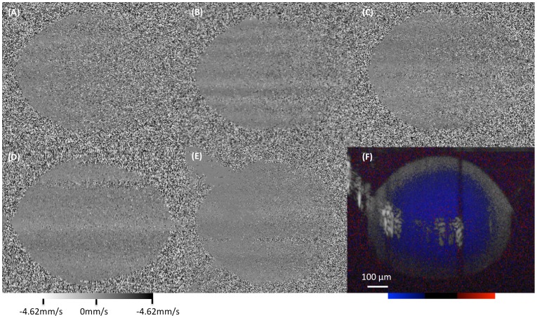
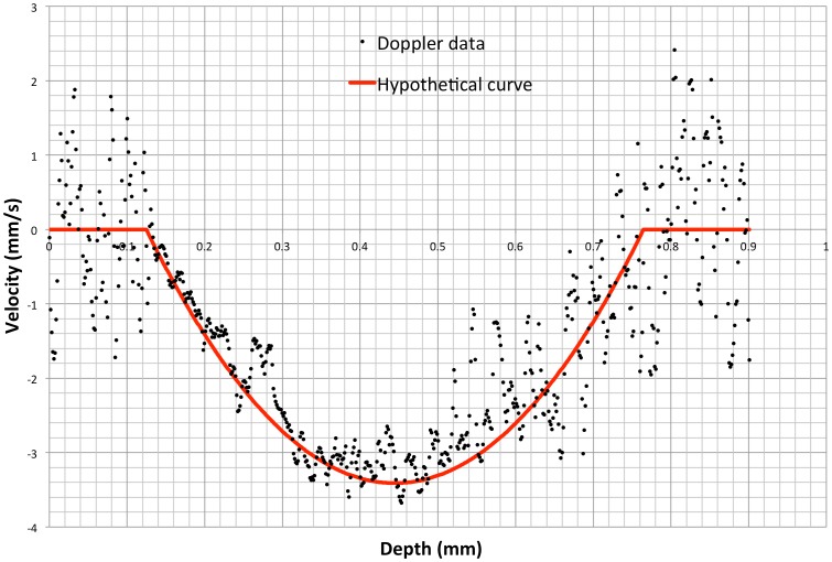
At the GNR concentration of 0.7×10(12) GNRs/mL, Doppler signal was present through the entire depth of the testing tube without substantial attenuation. A well-defined laminar flow profile was observed for Doppler images of GNRs flowing through the glass capillary tube. The Doppler OCT measured flow profile was not statistically different from the expected flow profile based upon an autoregressive moving average model, with an error of -0.025 to 0.037 mm/s (p = 0.6435). Cross-sectional slices demonstrated the ability to view anterior chamber outflow ex-vivo using GNR-enhanced Doppler OCT. Doppler volumetric flow measurements were comparable to flow recorded by the perfusion system.
GNRs created a measureable Doppler signal within otherwise silent flow fields in OCT Doppler scans. Practical application of this technique was confirmed in a constant pressure ex-vivo aqueous humor outflow model in porcine eyes.
研究金纳米棒(GNRs)作为一种造影剂,以增强巩膜内房水流出的多普勒光学相干断层扫描(OCT)成像。
用光谱域OCT设备(Bioptigen,北卡罗来纳州达勒姆)对GNRs的系列稀释液进行扫描,以可视化多普勒信号。使用可控流动系统对使用GNRs的多普勒测量进行验证。为了证明GNR增强多普勒的应用,对猪眼分别以恒压灌注单纯模拟房水或与模拟房水混合的1.0×10¹²个GNRs/mL。从角膜缘以30°增量的径向方式获得12次多普勒和容积SD-OCT扫描,形成圆形扫描模式。通过对角膜缘周围所有12次扫描中未连接血管内的血流进行积分来计算容积流量。
在GNR浓度为0.7×10¹²个GNRs/mL时,在整个测试管深度均存在多普勒信号,且无明显衰减。观察到GNRs流经玻璃毛细管的多普勒图像呈现出明确的层流轮廓。基于自回归移动平均模型,多普勒OCT测量的血流轮廓与预期血流轮廓在统计学上无差异,误差为-0.025至0.037 mm/s(p = 0.6435)。横截面切片显示,使用GNR增强多普勒OCT能够在体外观察前房流出情况。多普勒容积流量测量结果与灌注系统记录的流量相当。
在OCT多普勒扫描中,GNRs在原本无声的流场中产生了可测量的多普勒信号。该技术在猪眼恒压体外房水流出模型中得到了实际应用的证实。