Zamani Saeed, Hashemibeni Batool, Esfandiari Ebrahim, Kabiri Azadeh, Rabbani Hossein, Abutorabi Roshanak
Department of Anatomical Sciences and Molecular Biology, Medical Faculty, Isfahan University of Medical Sciences, Iran.
Department of Biophysics, Medical Faculty, Isfahan University of Medical Sciences, Iran.
Adv Biomed Res. 2014 Jan 27;3:54. doi: 10.4103/2277-9175.125799. eCollection 2014.
The Autologous Chondrocytes Transplantation (ACT) method is being studied for repair of cartilage diseases. As the chondrocytes dedifferentiated during monolayer culture, three-dimensional cultures are suggested to redifferentiate them. The aim of this study was investigation of the effect of TGF-β3 growth factor on chondrocytes in pellet culture system.
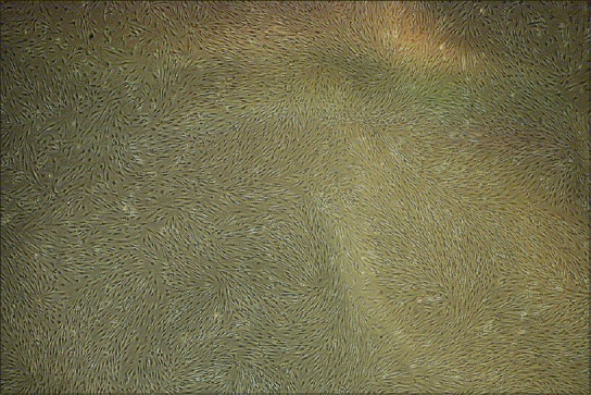
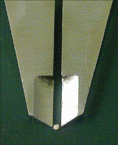
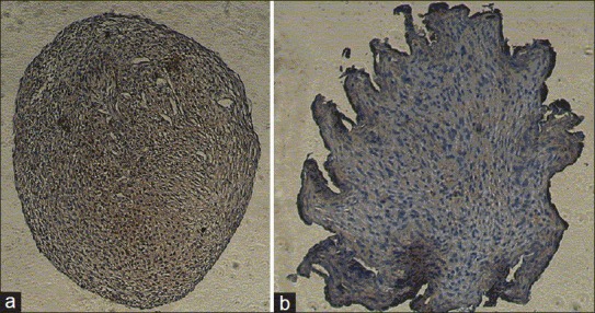
The chondrocytes were isolated from three human articular cartilages by enzymatic digestion. The cells of the second passage were transferred to pellet culture system. We determined the chondrogenic medium with TGF-β3 as the experimental group and without it as the control group. After 2 weeks, the aggrecan production was investigated using histological and immunohistochemical (IHC) methods.
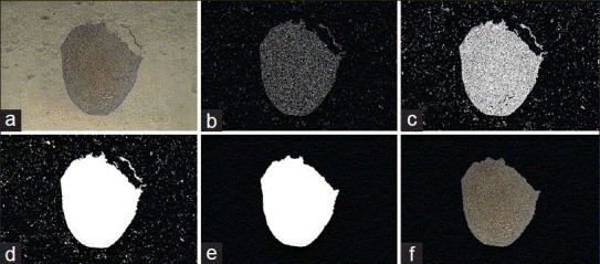
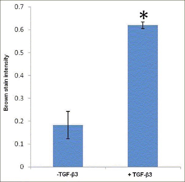
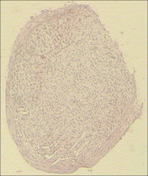
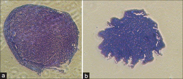
The presence of glycosaminoglycans was proved through Toluiden blue staining. Comparison of IHC results using MATLAB software showed that aggrecan in the experimental group was significantly higher than in the control group (P ≤ 0.05).
The presence of TGF-β3 in the chondrogenic medium could lead to the production of more aggrecan in chondrocytes cultivated in pellet culture system.
自体软骨细胞移植(ACT)方法正在用于软骨疾病修复的研究。由于软骨细胞在单层培养过程中会去分化,因此建议采用三维培养使其重新分化。本研究的目的是调查转化生长因子-β3(TGF-β3)生长因子对微团培养系统中软骨细胞的影响。
通过酶消化法从三块人类关节软骨中分离软骨细胞。将第二代细胞转移至微团培养系统。我们将添加TGF-β3的软骨形成培养基作为实验组,不添加TGF-β3的作为对照组。2周后,采用组织学和免疫组织化学(IHC)方法研究聚集蛋白聚糖的产生情况。
通过甲苯胺蓝染色证实了糖胺聚糖的存在。使用MATLAB软件对免疫组织化学结果进行比较显示,实验组中的聚集蛋白聚糖明显高于对照组(P≤0.05)。
软骨形成培养基中TGF-β3的存在可导致在微团培养系统中培养的软骨细胞产生更多的聚集蛋白聚糖。