Department of Chemistry, University of North Carolina, Chapel Hill, NC 27599, USA.
Lab Chip. 2014 May 7;14(9):1622-31. doi: 10.1039/c3lc51353j. Epub 2014 Mar 20.
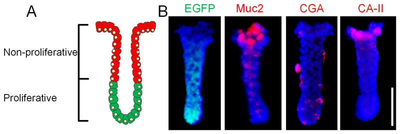
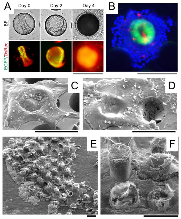
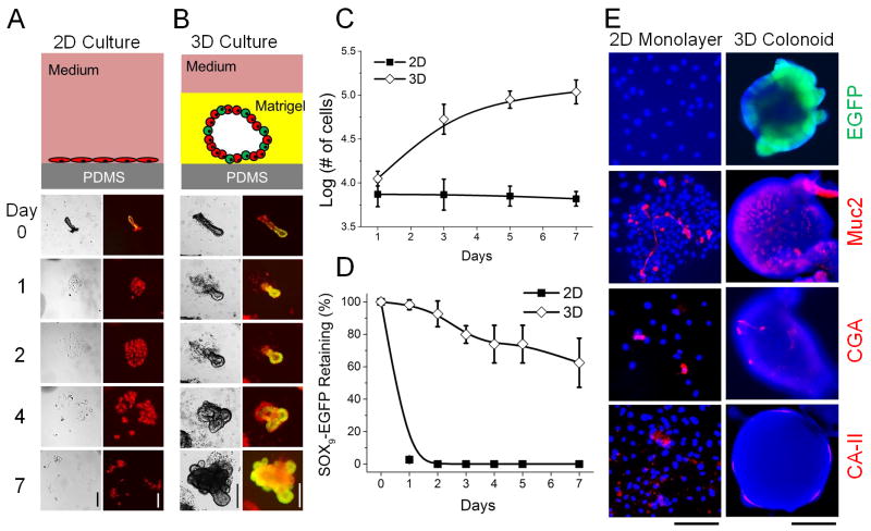
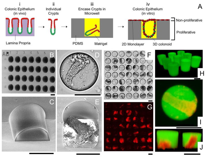
The proliferative compartment of the colonic epithelium in vivo is located in the basal crypt where colonic stem cells and transit-amplifying cells reside and fuel the rapid renewal of non-proliferative epithelial cells as they migrate toward the gut lumen. To mimic this tissue polarity, microstructures composed of polydimethylsiloxane (PDMS) microwells and Matrigel micropockets were used to guide a combined 2-dimensional (2D) and 3-dimensional (3D) hybrid culture of primary crypts isolated from the murine colon. The 2D and 3D culture of crypts on a planar PDMS surface was first investigated in terms of cell proliferation and stem cell activity. 3D culture of crypts with overlaid Matrigel generated enclosed, but highly proliferative spheroids (termed colonoids). 2D culture of crypts produced a spreading monolayer of cells, which were non-proliferative. A combined 2D/3D hybrid culture was generated in a PDMS microwell platform on which crypts were loaded by centrifugation into microwells (diameter = 150 μm, depth = 150 μm) followed by addition of Matrigel that formed micropockets locking the crypts within the microwells. Embedded crypts first underwent 3D expansion inside the wells. After the cells filled the microwells, they migrated onto the surrounding surface forming a 2D monolayer in the array regions without Matrigel. This unique 2D/3D hybrid culture generated a continuous, millimeter-scale colonic epithelial tissue in vitro, which resembled the polarized architecture (i.e. distinct proliferative and non-proliferative zones) and geometry of the colonic epithelium in vivo. This work initiates the construction of a "colon-on-a-chip" using primary cells/tissues with the ultimate goal of producing the physiologic structure and organ-level function of the colon.
体内结肠上皮的增殖区位于基底隐窝中,其中包含结肠干细胞和过渡扩增细胞,它们为非增殖性上皮细胞的快速更新提供动力,这些细胞在向肠腔迁移的过程中不断增殖。为了模拟这种组织极性,我们使用由聚二甲基硅氧烷(PDMS)微井和 Matrigel 微囊组成的微结构来引导从小鼠结肠中分离的原代隐窝的 2 维(2D)和 3 维(3D)混合培养。首先在 PDMS 平面上研究了隐窝的 2D 和 3D 培养,以评估细胞增殖和干细胞活性。在覆盖有 Matrigel 的情况下,3D 培养隐窝会产生封闭但高度增殖的球体(称为类器官)。2D 培养隐窝会产生铺展的单层细胞,这些细胞是非增殖性的。在 PDMS 微井平台上生成了一种结合的 2D/3D 混合培养,该平台通过离心将隐窝加载到微井(直径= 150μm,深度= 150μm)中,然后添加 Matrigel 形成微囊,将隐窝锁定在微井内。嵌入的隐窝首先在井内进行 3D 扩展。当细胞充满微井后,它们迁移到周围的表面上,在没有 Matrigel 的阵列区域中形成 2D 单层。这种独特的 2D/3D 混合培养在体外生成了连续的、毫米级的结肠上皮组织,该组织类似于体内结肠上皮的极化结构(即明显的增殖区和非增殖区)和几何形状。这项工作开创了使用原代细胞/组织构建“芯片上的结肠”的先河,最终目标是产生结肠的生理结构和器官水平功能。