Jiangsu Key Laboratory of Molecular and Functional Imaging, Department of Radiology, Zhongda Hospital, Medical School, Southeast University, Nanjing, China.
Department of Imaging and Interventional Radiology, Prince of Wales Hospital, the Chinese University of Hong Kong, Shatin, Hong Kong SAR, China.
PLoS One. 2014 Mar 25;9(3):e93124. doi: 10.1371/journal.pone.0093124. eCollection 2014.
Our purpose was to validate an early enhancement time point for accurately measuring the myocardial contrast partition coefficient (lambda) using dynamic-equilibrium magnetic resonance imaging.
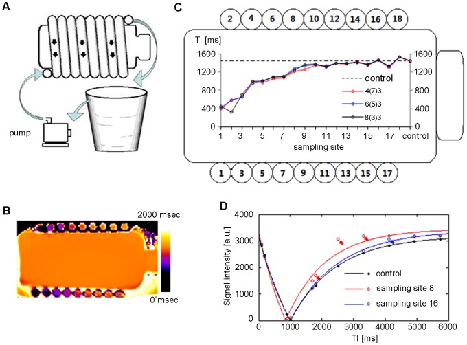
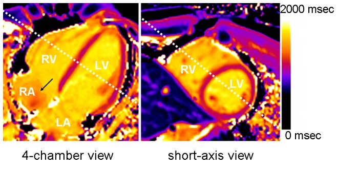
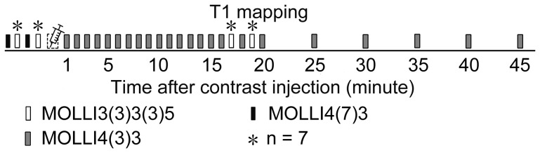
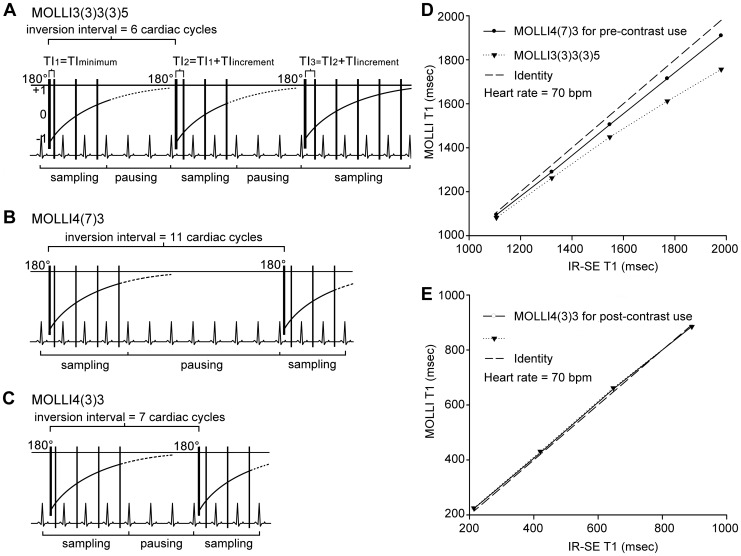
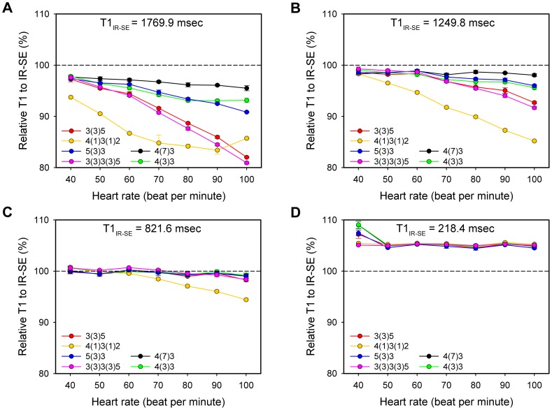
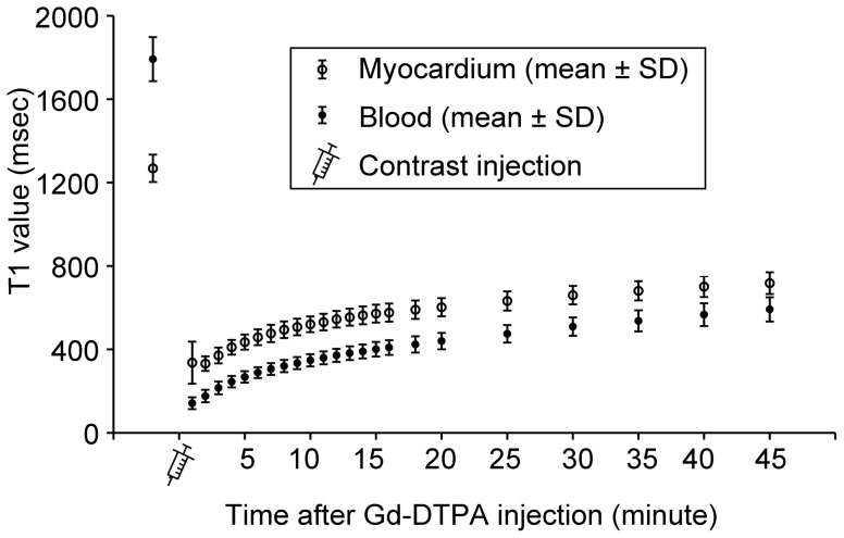
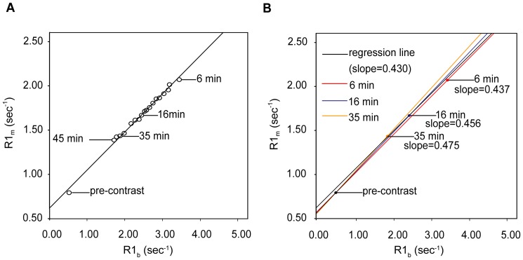
The pre- and post-contrast longitudinal relaxation rates (reciprocal of T1) of the interventricular septum (R1(m)) and blood pool (R1(b)) were obtained from fifteen healthy volunteers and three diabetic patients with hypertension using two optimized T1 mapping sequences (modified Look-Locker inversion recovery) on a 3-Tesla magnetic resonance scanner. Reference lambda values were calculated as the slope of the regression line of R1(m) versus R1(b) at dynamic equilibrium (multi-point regression method). The simplified pre-/post-enhancement two-acquisition method (two-point method) was used to calculate lambda by relating the change in R1(m) and R1(b) using different protocols according to the acquisition stage of the post-enhancement data point. The agreement with the referential method was tested by calculating Pearson's correlation coefficient and the intra-class correlation coefficient.
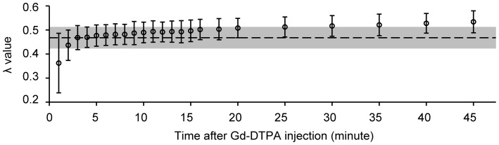
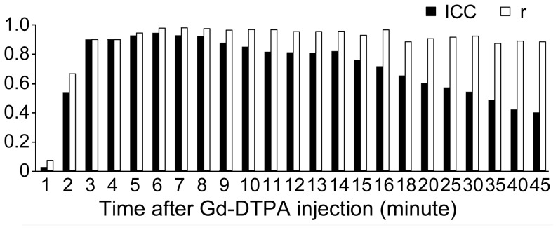
The lambda values measured by the two-point method increased (from 0.479 ± 0.041 to 0.534 ± 0.043) over time from 6 to 45 minutes after contrast and exhibited good correlation with the reference at each time point (r ≥ 0.875, p<0.05). The intra-class correlation coefficient on absolute agreement with the reference lambda was 0.946, 0.929 and 0.922 at the 6th, 7th and 8th minutes and dropped from 0.878 to 0.403 from the 9th minute on.
The time-efficient two-point method at 6-8 minutes after the Gd-DTPA bolus injection exhibited good agreement with the multi-point regression method and can be applied for accurate lambda measurement in normal myocardium.
本研究旨在验证一种新的早期强化时间点,以准确测量使用动态平衡磁共振成像的心肌对比分割系数(λ)。
15 名健康志愿者和 3 名高血压合并糖尿病患者在 3T 磁共振扫描仪上使用两种优化的 T1 映射序列(改良 Look-Locker 反转恢复)获得室间隔(R1(m))和血池(R1(b))的预对比和后对比纵向弛豫率(T1 的倒数)。参考λ值通过在动态平衡时(多点回归法)R1(m)与 R1(b)的回归线斜率计算得出。简化的预/后增强两采集法(两点法)用于通过根据后增强数据点采集阶段的不同协议,用 R1(m)和 R1(b)的变化来计算λ。用 Pearson 相关系数和组内相关系数检验与参考方法的一致性。
两点法测量的λ值从注射对比剂后 6 分钟到 45 分钟逐渐增加(从 0.479±0.041 增加到 0.534±0.043),并与每个时间点的参考值具有良好的相关性(r≥0.875,p<0.05)。与参考λ值的绝对一致性的组内相关系数在 6、7 和 8 分钟时分别为 0.946、0.929 和 0.922,从第 9 分钟开始降至 0.878 至 0.403。
Gd-DTPA 团注后 6-8 分钟时,这种时间效率高的两点法与多点回归法具有良好的一致性,可以用于正常心肌的准确λ测量。