Department of Radiology and Imaging Sciences, Indiana University School of Medicine, Indianapolis, Indiana, 46202; Stark Neurosciences Research Institute, Indiana University School of Medicine, Indianapolis, Indiana, 46202.
Synapse. 2014 Jun;68(6):266-74. doi: 10.1002/syn.21736. Epub 2014 Feb 28.
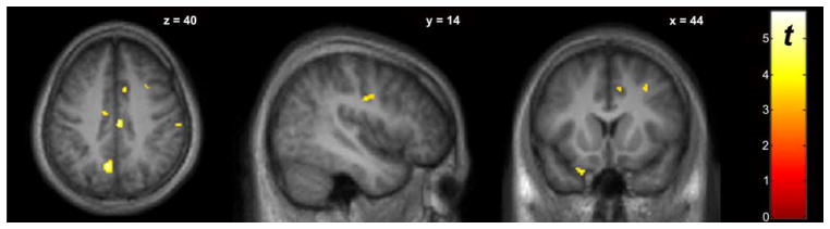
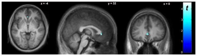
Dopamine (DA) dysregulation within fronto-striatal circuitry may underlie impulsivity in alcohol and other substance use disorders. To date, no one has directly demonstrated DA release during a task requiring the control of impulsive behavior. The current study was conducted to determine whether a response inhibition task (stop signal task; SST) would elicit detectable extrastriatal DA release in healthy controls. We hypothesized that DA release would be detected in regions previously implicated in different aspects of inhibitory control. [(18) F]Fallypride (FAL) PET imaging was performed in nine healthy males (24.6 ± 4.1 y.o.) to assess changes in cortical DA during a SST relative to a baseline "Go" task. On separate days, subjects received one FAL scan during the SST, and one FAL scan during a "Go" control; task-order was counter-balanced across subjects. Parametric BPND images were generated and analyzed with SPM8. Voxel-wise analysis indicated significant SST-induced DA release in several cortical regions involved in inhibitory control, including the insula, cingulate cortex, orbitofrontal cortex, precuneus, and supplementary motor area. There was a significant positive correlation between stop signal reaction time and DA release in the left orbitofrontal cortex, right middle frontal gyrus, and right precentral gyrus. These data support the feasibility of using FAL PET to study DA release during response inhibition, enabling investigation of relationships between DA function and impulsive behavior.
额纹体纹状体电路中的多巴胺(DA)失调可能是导致酒精和其他物质使用障碍中冲动的基础。迄今为止,没有人在需要控制冲动行为的任务中直接证明 DA 的释放。本研究旨在确定一项反应抑制任务(停止信号任务;SST)是否会在健康对照者中引起可检测的额纹体外 DA 释放。我们假设在先前涉及抑制控制不同方面的区域中会检测到 DA 释放。[(18)F]Fallypride(FAL)PET 成像在 9 名健康男性(24.6±4.1 岁)中进行,以评估相对于基线“Go”任务的 SST 期间皮质 DA 的变化。在不同的日子里,受试者在 SST 期间接受一次 FAL 扫描,在“Go”对照期间接受一次 FAL 扫描;任务顺序在受试者之间平衡。使用 SPM8 生成和分析参数 BPND 图像。体素分析表明,在涉及抑制控制的几个皮质区域中,包括岛叶、扣带皮层、眶额皮层、楔前叶和辅助运动区,SST 诱导的 DA 释放显著。左眶额皮质、右中央前回和右中央前回的 DA 释放与停止信号反应时间呈显著正相关。这些数据支持使用 FAL PET 研究反应抑制期间 DA 释放的可行性,从而能够研究 DA 功能与冲动行为之间的关系。