Department of Pediatrics, Vanderbilt University, Nashville, TN, USA.
BMC Pediatr. 2014 Mar 28;14:84. doi: 10.1186/1471-2431-14-84.
Magnetic resonance imaging (MRI) is a useful tool to study brain growth and organization in preterm neonates for clinical and research purposes, but its practicality can be limited by time and medical constraints. The aim of this study was to determine if MRI relaxometry of the deep nuclei, as opposed to white matter, would reflect the influence of gestational age at birth on structures essential to motor development, regardless of postnatal age at the time of imaging.
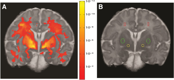
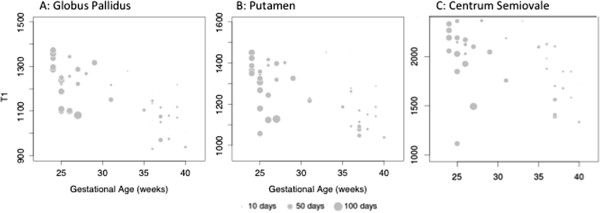
This was a prospective observational study of infants without brain injury on conventional neuroimaging who were cared for in the neonatal intensive care unit (NICU) at Vanderbilt. Infants were studied using MRI relaxometry within a 2-month window of postmenstrual term age. In 45 infants, white matter MRI T1 relaxation times were influenced by both gestational age and postnatal age at imaging time (R(2) = 0.19 for gestational age vs. R(2) = 0.34 adjusting for both gestational age and age at imaging; all p < 0.01). Similar results were obtained with T2 relaxation times. In contrast, globus pallidus T1 reflected gestational age but was minimally affected by postnatal age (R(2) = 0.50 vs. 0.57, p < 0.001).
The results obtained using this imaging protocol are consistent with the slow maturation of the globus pallidus, essential to normal development of complex motor programs into adulthood. Globus pallidus MRI relaxometry measures the impact of gestational age at birth on brain development independent of postnatal age in preterm infants and should prove useful for predictive modeling in a flexible time-window around postmenstrual term age.
磁共振成像(MRI)是一种用于研究早产儿脑生长和组织的有用工具,可用于临床和研究目的,但由于时间和医疗限制,其实际应用可能受到限制。本研究旨在确定与白质相比,深部核的 MRI 弛豫度是否反映了出生时的胎龄对运动发育至关重要的结构的影响,而与成像时的产后年龄无关。
这是一项前瞻性观察性研究,研究对象为在范德比尔特新生儿重症监护病房(NICU)接受常规神经影像学检查且无脑损伤的婴儿。在出生后平均年龄的 2 个月窗口期内,使用 MRI 弛豫度对婴儿进行研究。在 45 名婴儿中,白质 MRI T1 弛豫时间既受胎龄又受成像时的产后年龄影响(胎龄的 R²=0.19,调整胎龄和成像时年龄后的 R²=0.34;所有 p<0.01)。T2 弛豫时间也得到了类似的结果。相比之下,苍白球 T1 反映胎龄,但受产后年龄影响较小(R²=0.50 与 0.57,p<0.001)。
使用该成像方案获得的结果与苍白球的缓慢成熟一致,这对成年期正常发育复杂运动程序至关重要。苍白球 MRI 弛豫度测量出生时胎龄对早产儿脑发育的影响,与产后年龄无关,在出生后平均年龄的灵活时间窗口内,应有助于预测建模。