Department of Cell Biology, State University of New York Downstate Medical Center, Brooklyn, New York; Neural and Behavioral Science Graduate Program, State University of New York Downstate Medical Center, Brooklyn, New York.
Glia. 2014 Jul;62(7):1053-65. doi: 10.1002/glia.22661. Epub 2014 Mar 31.
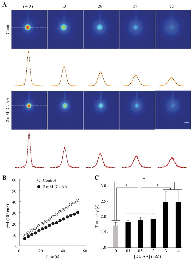
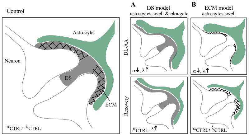
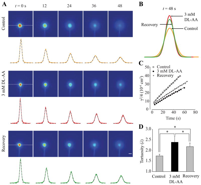
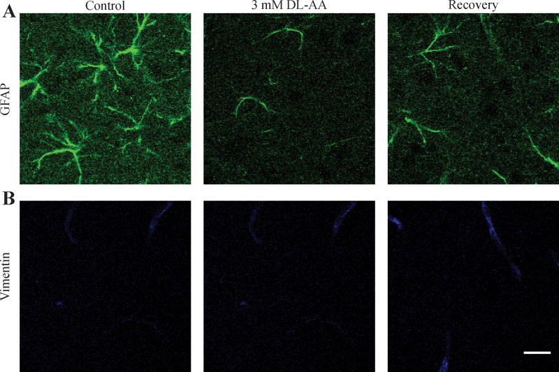
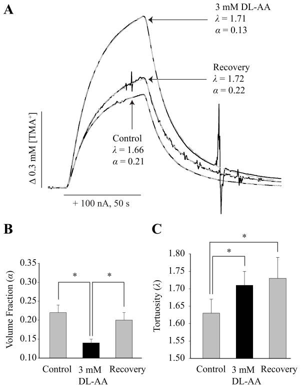
One of the hallmarks of numerous life-threatening and debilitating brain diseases is cellular swelling that negatively impacts extracellular space (ECS) structure. The ECS structure is determined by two macroscopic parameters, namely tortuosity (λ) and volume fraction (α). Tortuosity represents hindrance imposed on the diffusing molecules by the tissue in comparison with an obstacle-free medium. Volume fraction is the proportion of tissue volume occupied by the ECS. From a clinical perspective, it is essential to recognize which factors determine the ECS parameters and how these factors change in brain diseases. Previous studies demonstrated that dead-space (DS) microdomains increased λ during ischemia and hypotonic stress, as these pocket-like structures transiently trapped diffusing molecules. We hypothesize that astrocytes play a key role in the formation of DS microdomains because their thin processes have concave shapes that may elongate as astrocytes swell in these pathologies. Here we selectively swelled astrocytes in the somatosensory neocortex of rat brain slices with a gliotoxin DL-α-Aminoadipic Acid (DL-AA), and we quantified the ECS parameters using Integrative Optical Imaging (IOI) and Real-Time Iontophoretic (RTI) diffusion methods. We found that α decreased and λ increased during DL-AA application. During recovery, α was restored whereas λ remained elevated. Increase in λ during astrocytic swelling and recovery is consistent with the formation of DS microdomains. Our data attribute to the astrocytes an important role in determining the ECS parameters, and indicate that extracellular diffusion can be improved not only by reducing the swelling but also by disrupting the DS microdomains.
许多危及生命和使人衰弱的脑部疾病的一个特征是细胞肿胀,这会对细胞外空间 (ECS) 的结构产生负面影响。ECS 的结构由两个宏观参数决定,即迂曲度 (λ) 和体积分数 (α)。迂曲度代表与无阻碍介质相比,组织对扩散分子造成的阻碍。体积分数是 ECS 占据组织体积的比例。从临床角度来看,识别哪些因素决定了 ECS 参数以及这些因素在脑部疾病中如何变化是至关重要的。先前的研究表明,在缺血和低渗应激期间,死腔 (DS) 微域会增加 λ,因为这些袋状结构会暂时困住扩散分子。我们假设星形胶质细胞在 DS 微域的形成中发挥关键作用,因为它们的薄突具有凹形,当星形胶质细胞在这些病变中肿胀时,这些凹形可能会延长。在这里,我们使用整合光学成像 (IOI) 和实时离子电泳 (RTI) 扩散方法,选择性地使大鼠大脑皮层感觉区的星形胶质细胞肿胀,并使用上述方法定量 ECS 参数。我们发现,在 DL-AA 应用过程中,α 降低,λ 增加。在恢复过程中,α 得到恢复,而 λ 仍然升高。在星形胶质细胞肿胀和恢复过程中 λ 的增加与 DS 微域的形成一致。我们的数据表明星形胶质细胞在确定 ECS 参数方面起着重要作用,并表明不仅通过减少肿胀,而且通过破坏 DS 微域,细胞外扩散可以得到改善。