Vallés Ana S, Aveldaño Marta I, Furland Natalia E
Instituto de Investigaciones Bioquímicas de Bahía Blanca, Consejo Nacional de Investigaciones Científicas y Técnicas (CONICET) y Universidad Nacional del Sur (UNS), Bahía Blanca, Argentina.
PLoS One. 2014 Apr 1;9(4):e91127. doi: 10.1371/journal.pone.0091127. eCollection 2014.
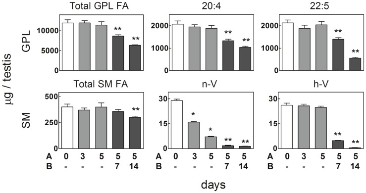
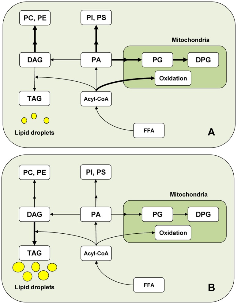
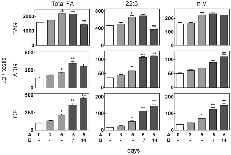
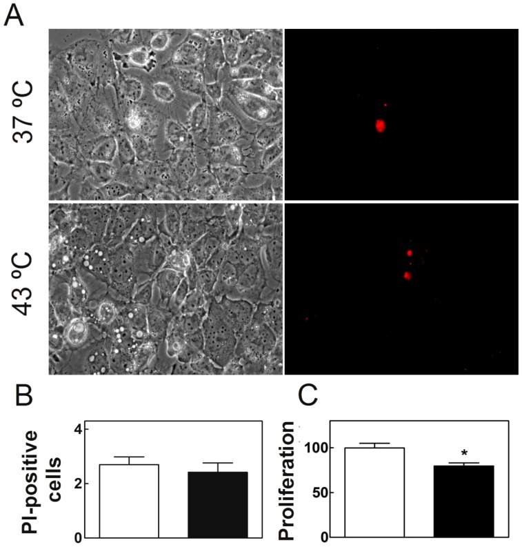
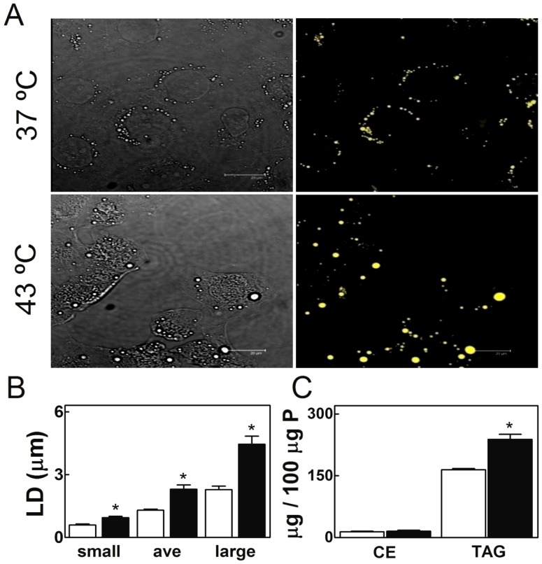
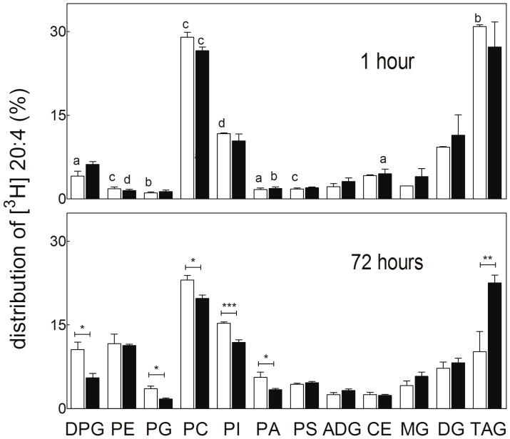
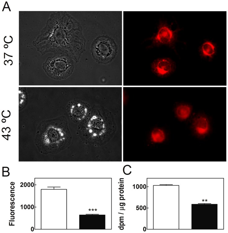
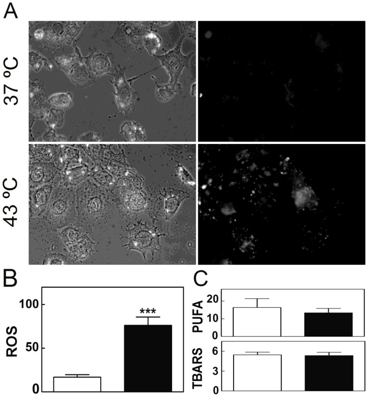
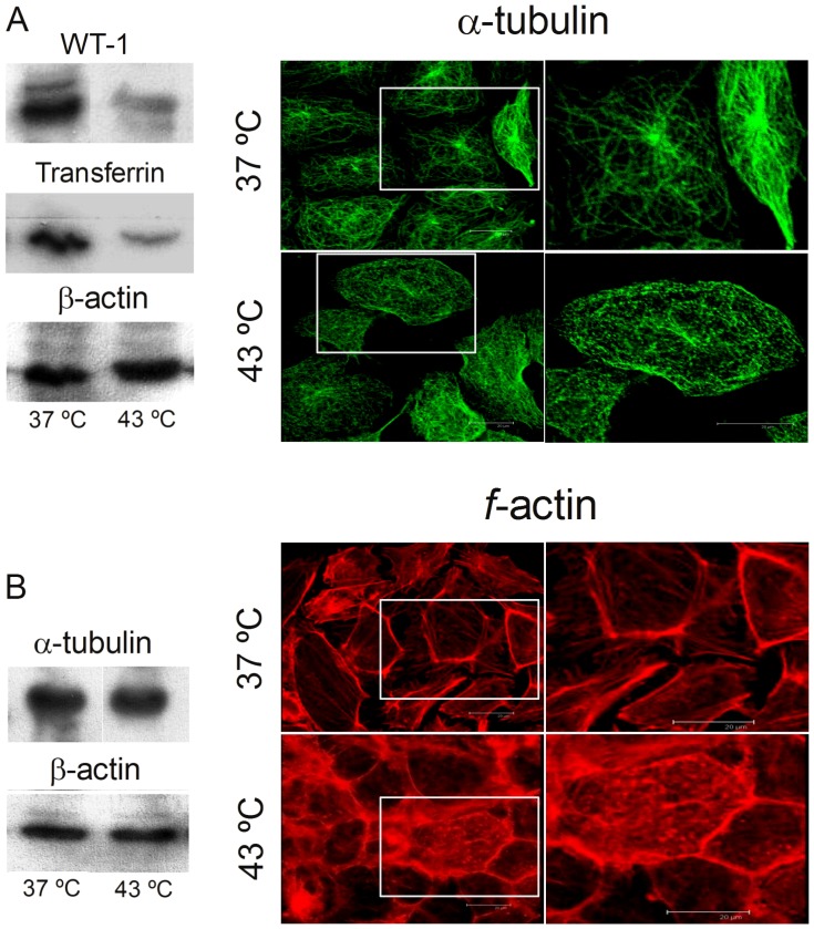
Spermatogenesis is known to be vulnerable to temperature. Exposures of rat testis to moderate hyperthermia result in loss of germ cells with survival of Sertoli cells (SC). Because SC provide structural and metabolic support to germ cells, our aim was to test the hypothesis that these exposures affect SC functions, thus contributing to germ cell damage. In vivo, regularly repeated exposures (one of 15 min per day, once a day during 5 days) of rat testes to 43 °C led to accumulation of neutral lipids. This SC-specific lipid function took 1-2 weeks after the last of these exposures to be maximal. In cultured SC, similar daily exposures for 15 min to 43 °C resulted in significant increase in triacylglycerol levels and accumulation of lipid droplets. After incubations with [3H]arachidonate, the labeling of cardiolipin decreased more than that of other lipid classes. Another specifically mitochondrial lipid metabolic function, fatty acid oxidation, also declined. These lipid changes suggested that temperature affects SC mitochondrial physiology, which was confirmed by significantly increased degrees of membrane depolarization and ROS production. This concurred with reduced expression of two SC-specific proteins, transferrin, and Wilms' Tumor 1 protein, markers of SC secretion and differentiation functions, respectively, and with an intense SC cytoskeletal perturbation, evident by loss of microtubule network (α-tubulin) and microfilament (f-actin) organization. Albeit temporary and potentially reversible, hyperthermia-induced SC structural and metabolic alterations may be long-lasting and/or extensive enough to respond for the decreased survival of the germ cells they normally foster.
已知精子发生对温度敏感。将大鼠睾丸暴露于适度高温会导致生殖细胞损失,而支持细胞(SC)存活。由于支持细胞为生殖细胞提供结构和代谢支持,我们的目的是检验以下假设:这些暴露会影响支持细胞的功能,从而导致生殖细胞损伤。在体内,将大鼠睾丸定期重复暴露(每天15分钟,共5天)于43°C会导致中性脂质积累。这种支持细胞特异性的脂质功能在最后一次暴露后1 - 2周达到最大值。在培养的支持细胞中,每天类似地暴露于43°C 15分钟会导致三酰甘油水平显著增加和脂滴积累。用[3H]花生四烯酸孵育后,心磷脂的标记下降幅度大于其他脂质类别。另一种特异性的线粒体脂质代谢功能,即脂肪酸氧化,也下降了。这些脂质变化表明温度影响支持细胞的线粒体生理,这通过膜去极化程度和活性氧产生的显著增加得到证实。这与两种支持细胞特异性蛋白,转铁蛋白和威尔姆斯瘤1蛋白的表达降低相一致,它们分别是支持细胞分泌和分化功能的标志物,并且伴随着明显的支持细胞细胞骨架扰动,表现为微管网络(α - 微管蛋白)和微丝(F - 肌动蛋白)组织的丧失。尽管高温诱导的支持细胞结构和代谢改变是暂时的且可能可逆,但可能持续时间足够长和/或范围足够广泛,足以导致它们正常培养的生殖细胞存活率下降。