Abdel Aziz Mt, Atta Hm, Roshdy Nk, Rashed LA, Sabry D, Hassouna Aa, Aboul Fotouh Gi, Hasan Nm, Younis Rh, Chowdhury Jr
Departments of Medical Biochemistry, Unit of Biochemistry and Molecular Biology and Histology .
Faculty of Medicine, Cairo University , Cairo, Egypt .
J Stem Cells Regen Med. 2012 Apr 14;8(1):28-34. doi: 10.46582/jsrm.0801005. eCollection 2012.
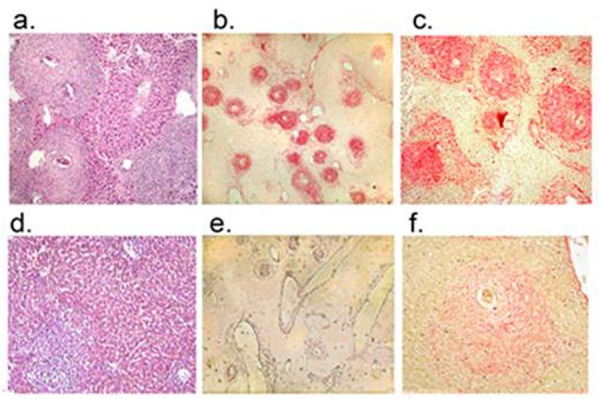
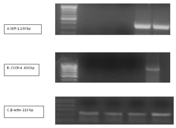
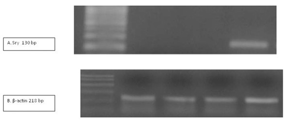
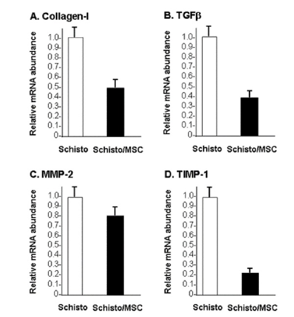
Schistosomiasis is a common chronic helminthic infection of the liver that causes hepatic fibrosis and portal hypertension,contributing to the death of over half a million people a year. Infusion of autologous bone marrow cells into patients with hepatic cirrhosis has been reported to ameliorate symptoms of portal hypertension and improve liver function, either by conversion of the infused mesenchymal stem cells (MSCs) to hepatocytes or by modulating of the hepatic fibrosis. Here,we have investigated the antifibrotic effect of mesenchymal stem cells (MSCs) using S. mansoni-induced liver fibrosis in mice, which causes an intense, stable fibrosis. MSCs derived from bone marrow of male mice were then infused intravenously into female mice that had received intraperitoneal injection of S.mansoni cercariae. Mice were divided into 4 groups: Untreated control; MSCs infusion only; Schistosomiasis only; and Schistosomiasis plus MSCs infusion. Serum alanine aminotransferase (ALT) and liver histopathology were evaluated. Expression of the collagen gene (type I),transforming growth factor (TGF-β), matrix metalloproteinase (MMP2), tissue inhibitor of metalloproteinase (TIMP-1),stromal cell-derived factor-1(SDF-1) and its receptor (CXCR4) were analyzed. MSC infusion resulted in significant decrease in liver collagen and TGF-β gene expression in the Schistosomiasis mice. The ratio of MMP-2 to TIMP-1 expression increased. SDF-1 and CXCR4 mRNA expression also increased. There was overall improvement of liver histology and a statistically significant reduction of serum ALT level. MSCs infusion ameliorated S. mansoni-induced liver fibrosis, probably by modulating the relative expression of MMP and TIMP. The findings support the hypothesis that MSCs participate in liver regeneration and functional improvement by reducing liver fibrosis.
血吸虫病是一种常见的肝脏慢性蠕虫感染,可导致肝纤维化和门静脉高压,每年造成超过50万人死亡。据报道,向肝硬化患者输注自体骨髓细胞可改善门静脉高压症状并改善肝功能,这可能是通过将输注的间充质干细胞(MSC)转化为肝细胞,或者通过调节肝纤维化来实现的。在此,我们利用曼氏血吸虫诱导的小鼠肝纤维化模型(该模型可导致强烈且稳定的纤维化),研究了间充质干细胞(MSC)的抗纤维化作用。然后将源自雄性小鼠骨髓的MSC静脉内输注到已接受腹腔注射曼氏血吸虫尾蚴的雌性小鼠体内。小鼠被分为4组:未治疗对照组;仅输注MSC组;仅患血吸虫病组;患血吸虫病加输注MSC组。评估了血清丙氨酸氨基转移酶(ALT)和肝脏组织病理学。分析了胶原蛋白基因(I型)、转化生长因子(TGF-β)、基质金属蛋白酶(MMP2)、金属蛋白酶组织抑制剂(TIMP-1)、基质细胞衍生因子-1(SDF-1)及其受体(CXCR4)的表达。在患血吸虫病的小鼠中,输注MSC导致肝脏胶原蛋白和TGF-β基因表达显著降低。MMP-2与TIMP-1表达的比值增加。SDF-1和CXCR4 mRNA表达也增加。肝脏组织学总体改善,血清ALT水平有统计学意义的降低。输注MSC改善了曼氏血吸虫诱导的肝纤维化,可能是通过调节MMP和TIMP的相对表达来实现的。这些发现支持了MSC通过减少肝纤维化参与肝脏再生和功能改善的假说。