Mazzuca Enrico, Salito Caterina, Rivolta Ilaria, Aliverti Andrea, Miserocchi Giuseppe
TBM Lab, Dipartimento di Elettronica, Informazione e Bioingegneria, Politecnico di Milano, Milano, Italy.
Department of Health Sciences, Università di Milano-Bicocca, Monza, Italy.
Physiol Rep. 2014 Feb 7;2(2):e00221. doi: 10.1002/phy2.221. eCollection 2014 Feb 1.
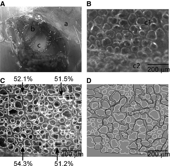
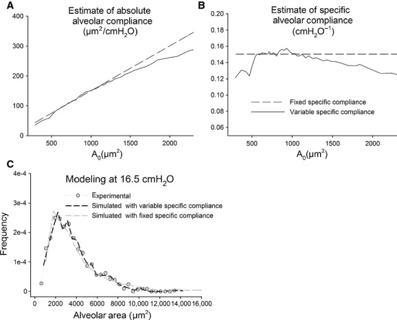
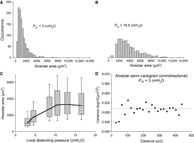
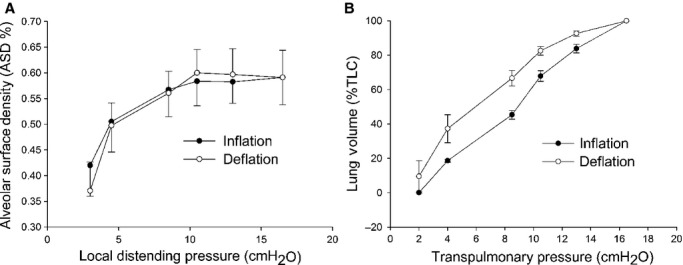
In six male anesthetized, tracheotomized, and mechanically ventilated rabbits, we imaged subpleural alveoli under microscopic view (60×) through a "pleural window" obtained by stripping the endothoracic fascia and leaving the parietal pleura intact. Three different imaging scale levels were identified for the analysis on increasing stepwise local distending pressure (P ld) up to 16.5 cmH2O: alveoli, alveolar cluster, and whole image field. Alveolar profiles were manually traced, clusters of alveoli of similar size were identified through a contiguity-constrained hierarchical agglomerative clustering analysis and alveolar surface density (ASD) was estimated as the percentage of air on the whole image field. Alveolar area distributions were remarkably right-skewed and showed an increase in median value with a large topology-independent heterogeneity on increasing P ld. Modeling of alveolar area distributions on increasing P ld led to hypothesize that absolute alveolar compliance (change in surface area over change in P ld) increases fairly linearly with increasing initial alveolar size, the corollary of this assumption being a constant specific compliance. Clusters were reciprocally interweaved due to their highly variable complex shapes. ASD was found to increase with a small coefficient of variation (CV <25%) with increasing P ld. The CV of lung volume at each transpulmonary pressure was further decreased (about 6%). The results of the study suggest that the considerable heterogeneity of alveolar size and of the corresponding alveolar mechanical behavior are homogenously distributed, resulting in a substantially homogenous mechanical behavior of lung units and whole organ.
在六只麻醉、气管切开并机械通气的雄性兔子中,我们通过剥离胸内筋膜并保持脏胸膜完整获得“胸膜窗”,在显微镜下(60倍)对胸膜下肺泡进行成像。确定了三种不同的成像尺度水平,用于逐步增加局部扩张压力(P ld)直至16.5 cmH₂O时的分析:肺泡、肺泡簇和整个图像视野。手动追踪肺泡轮廓,通过相邻约束层次凝聚聚类分析识别大小相似的肺泡簇,并将肺泡表面密度(ASD)估计为整个图像视野中空气的百分比。肺泡面积分布明显右偏,并且随着P ld的增加,中位数增加,且具有与拓扑无关的大异质性。对P ld增加时肺泡面积分布进行建模后推测,绝对肺泡顺应性(表面积变化与P ld变化之比)随着初始肺泡大小的增加而相当线性地增加,这一假设的必然结果是特定顺应性恒定。由于其高度可变的复杂形状,簇相互交织。发现ASD随着P ld的增加而以较小的变异系数(CV <25%)增加。每个跨肺压下肺容积的CV进一步降低(约6%)。研究结果表明,肺泡大小和相应肺泡力学行为的显著异质性均匀分布,导致肺单位和整个器官的力学行为基本均匀。