Lloyd Mark C, Alfarouk Khalid O, Verduzco Daniel, Bui Marilyn M, Gillies Robert J, Ibrahim Muntaser E, Brown Joel S, Gatenby Robert A
H, Lee Moffitt Cancer Center, 12902 Magnolia Drive, Tampa, FL 33612, USA.
BMC Cancer. 2014 Apr 23;14:279. doi: 10.1186/1471-2407-14-279.
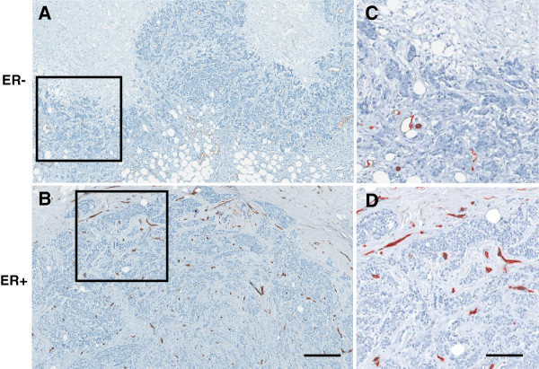
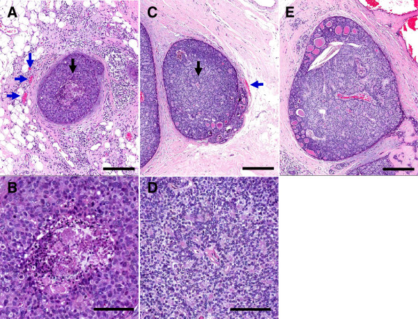
Breast carcinoma can be classified as either Estrogen Receptor (ER) positive or negative by immunohistochemical phenotyping, although ER expression may vary from 1 to 100% of malignant cells within an ER + tumor. This is similar to genetic variability observed in other tumor types and is generally viewed as a consequence of intratumoral evolution driven by random genetic mutations. Here we view cellular evolution within tumors as a classical Darwinian system in which variations in molecular properties represent predictable adaptations to spatially heterogeneous environmental selection forces. We hypothesize that ER expression is a successful adaptive strategy only if estrogen is present in the microenvironment. Since the dominant source of estrogen is blood flow, we hypothesized that, in general, intratumoral regions with higher blood flow would contain larger numbers of ER + cells when compared to areas of low blood flow and in turn necrosis.
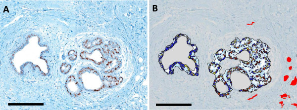
This study used digital pathology whole slide image acquisition and advanced image analysis algorithms. We examined the spatial distribution of ER + and ER- cells, vascular density, vessel area, and tissue necrosis within histological sections of 24 breast cancer specimens. These data were correlated with the patients ER status and molecular pathology report findings.
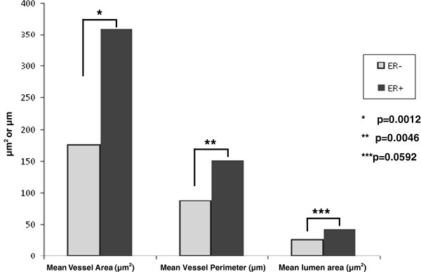
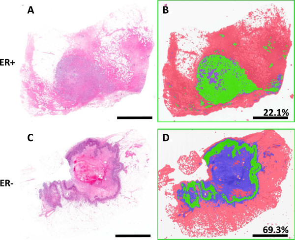
ANOVA analyses revealed a strong correlation between vascular area and ER expression and between high fractional necrosis and absent ER expression (R(2) = 39%; p < 0.003 and R(2) = 46%; p < 0.001), respectively). ER expression did not correlate with tumor grade or size.
We conclude that ER expression can be understood as a Darwinian process and linked to variations in estrogen delivery by temporal and spatial heterogeneity in blood flow. This correlation suggests strategies to promote intratumoral blood flow or a cyclic introduction of estrogen in the treatment schedule could be explored as a counter-intuitive approach to increase the efficacy of anti-estrogen drugs.
通过免疫组化表型分析,乳腺癌可分为雌激素受体(ER)阳性或阴性,尽管在ER +肿瘤中,ER表达在1%至100%的恶性细胞之间存在差异。这与在其他肿瘤类型中观察到的基因变异性相似,通常被视为由随机基因突变驱动的肿瘤内进化的结果。在这里,我们将肿瘤内的细胞进化视为一个经典的达尔文系统,其中分子特性的变化代表了对空间异质环境选择力的可预测适应。我们假设,只有当微环境中存在雌激素时,ER表达才是一种成功的适应性策略。由于雌激素的主要来源是血流,我们推测,一般来说,与低血流区域及坏死区域相比,血流较高的肿瘤内区域会含有更多的ER +细胞。
本研究使用了数字病理全玻片图像采集和先进的图像分析算法。我们检查了24个乳腺癌标本组织切片中ER +和ER -细胞的空间分布、血管密度、血管面积和组织坏死情况。这些数据与患者的ER状态和分子病理报告结果相关联。
方差分析显示,血管面积与ER表达之间以及高坏死分数与ER表达缺失之间分别存在强相关性(R(2) = 39%;p < 0.003和R(2) = 46%;p < 0.001)。ER表达与肿瘤分级或大小无关。
我们得出结论,ER表达可被理解为一个达尔文过程,并与血流的时空异质性导致的雌激素输送变化有关。这种相关性表明,作为一种增加抗雌激素药物疗效的反直觉方法,可以探索促进肿瘤内血流或在治疗方案中周期性引入雌激素的策略。