Kim Soo-Ah, Hong Ran
Department of Obstetrics and Gynecology, College of Medicine, Chosun University, Gwangju 501-759, Republic of Korea.
Department of Pathology, College of Medicine, Chosun University, Gwangju 501-759, Republic of Korea.
Oncol Lett. 2014 May;7(5):1589-1593. doi: 10.3892/ol.2014.1948. Epub 2014 Mar 7.
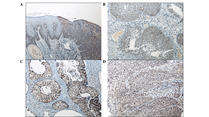
Survivin is a member of the inhibitor of apoptosis protein family. Under normal circumstances, survivin is expressed in embryonic and fetal tissues, but is completely downregulated in normal adult tissues. Notably, this protein has been found to be prominently expressed in a variety of human malignant tumors. The present study was designed to evaluate the possible role of survivin in the tumorigenesis of cervical intraepithelial neoplasia and invasive squamous cell carcinoma (SCC) of the uterine cervix. In addition, it was investigated whether the nuclear or cytoplasmic expression of survivin is associated with tumor progression. In total, 71 samples of cervical squamous tissue were obtained, including 15 normal squamous epithelia, 25 high-grade squamous intraepithelial lesions (HSILs) and 31 SCCs, from cone biopsy and hysterectomy specimens and stained for survivin expression by immunohistochemistry. The intensity of survivin expression tended to increase with tumor progression (60.0% of normal mucosa, 76.0% of HSIL and 80.6% of SCC samples demonstrated high intensity survivin expression), but this correlation was not found to be statistically significant. However, a statistically significant difference was identified in the intracellular localization of survivin among the normal mucosa, HSIL and SCC samples (P<0.001). In total, 72% (18/25) of HSIL and 54.8% (17/31) of SCC cases expressed cytoplasmic staining in contrast to the nuclear staining of the normal mucosa. In addition, 64% (16/25) of HSIL and 42% (13/31) of SCC cases showed coexpression in the nucleus and cytoplasm. An inverse correlation was identified between the decrement of nuclear survivin expression and tumor progression, but was not statistically significant (P=0.08). These results indicated that analysis of the intracellular expression of survivin (particularly cytoplasmic expression) is a marker for predicting disease progression in the uterine cervix.
生存素是凋亡抑制蛋白家族的成员。在正常情况下,生存素在胚胎和胎儿组织中表达,但在正常成人组织中完全下调。值得注意的是,已发现该蛋白在多种人类恶性肿瘤中显著表达。本研究旨在评估生存素在宫颈上皮内瘤变和子宫颈浸润性鳞状细胞癌(SCC)发生中的可能作用。此外,还研究了生存素的核表达或胞质表达是否与肿瘤进展相关。总共从锥形活检和子宫切除标本中获取了71份宫颈鳞状组织样本,包括15份正常鳞状上皮、25份高级别鳞状上皮内病变(HSIL)和31份SCC,并通过免疫组织化学对生存素表达进行染色。生存素表达强度倾向于随肿瘤进展而增加(60.0%的正常黏膜、76.0%的HSIL和80.6%的SCC样本显示高强度生存素表达),但未发现这种相关性具有统计学意义。然而,在正常黏膜、HSIL和SCC样本中,生存素的细胞内定位存在统计学显著差异(P<0.001)。与正常黏膜的核染色相比,总共72%(18/25)的HSIL和54.8%(17/31)的SCC病例表现为胞质染色。此外,64%(16/25)的HSIL和42%(13/31)的SCC病例显示核和胞质共表达。核生存素表达的减少与肿瘤进展呈负相关,但无统计学意义(P=0.08)。这些结果表明,分析生存素的细胞内表达(特别是胞质表达)是预测子宫颈疾病进展的一个标志物。