Marchesini Jlenia, Campo Gianluca, Righi Riccardo, Benea Giorgio, Ferrari Roberto
Cardiovascular Institute, Azienda Ospedaliero-Universitaria S. Anna, Ferrara and Cardiovascular Research Centre, Salvatore Maugeri Foundation, IRCCS, Lumezzane (BS) and LTTA Center, Ferrara;
U.O. di Diagnostica per immagini e Radiologia Interventistica, Ospedale del Delta, Lagosanto, Ferrara, Italy.
Clin Pract. 2011 Nov 8;1(4):e107. doi: 10.4081/cp.2011.e107. eCollection 2011 Sep 28.
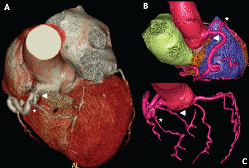
ST-segment elevation MI (STEMI) is a rare presentation in patients with coronary artery anomalies. In these patients, the identification of the culprit lesion and its treatment may be difficult, particularly in the emergency setting of primary percutaneous coronary intervention (PCI). From January 2008 to April 2011, 1015 STEMI patients received coronary artery angiography and primary PCI in our centre. Of these, 5 (0.4%) patients showed a coronary artery anomaly. In this paper we reported two rare cases: i) the first is a single coronary artery originating from right sinus of Valsalva; ii) the second is a separate origin of 3 coronary arteries originating from the right sinus of Valsalva. In conclusion, coronary artery anomalies presenting with STEMI are really uncommon, but often are a challenge. The integration between traditional coronary artery angiography and multidetector computerized tomography is crucial to optimize the interventional and medical management of these patients.
ST段抬高型心肌梗死(STEMI)在冠状动脉异常患者中是一种罕见的表现。在这些患者中,确定罪犯病变及其治疗可能很困难,尤其是在急诊行直接经皮冠状动脉介入治疗(PCI)的情况下。2008年1月至2011年4月,1015例STEMI患者在我们中心接受了冠状动脉造影和直接PCI。其中,5例(0.4%)患者表现为冠状动脉异常。在本文中,我们报告了两例罕见病例:i)第一例是一条起源于瓦尔萨尔瓦右窦的单一冠状动脉;ii)第二例是三条冠状动脉分别起源于瓦尔萨尔瓦右窦。总之,以STEMI为表现的冠状动脉异常确实不常见,但往往具有挑战性。传统冠状动脉造影与多排螺旋计算机断层扫描相结合对于优化这些患者的介入和药物治疗至关重要。