Bryant Nathan D, Li Ke, Does Mark D, Barnes Stephanie, Gochberg Daniel F, Yankeelov Thomas E, Park Jane H, Damon Bruce M
Vanderbilt University Institute of Imaging Science, Vanderbilt University, Nashville, TN, USA; Department of Radiology and Radiological Sciences, Vanderbilt University, Nashville, TN, USA.
NMR Biomed. 2014 Jun;27(6):716-25. doi: 10.1002/nbm.3113. Epub 2014 Apr 29.
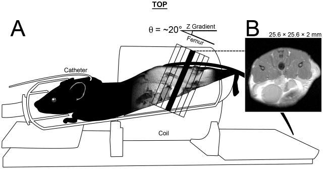
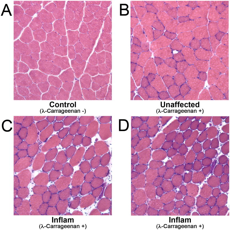
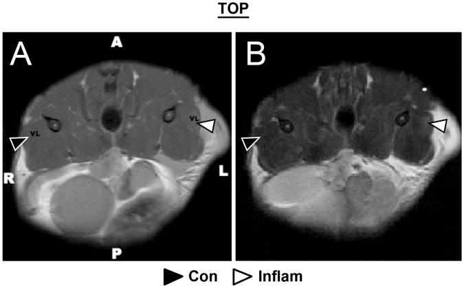
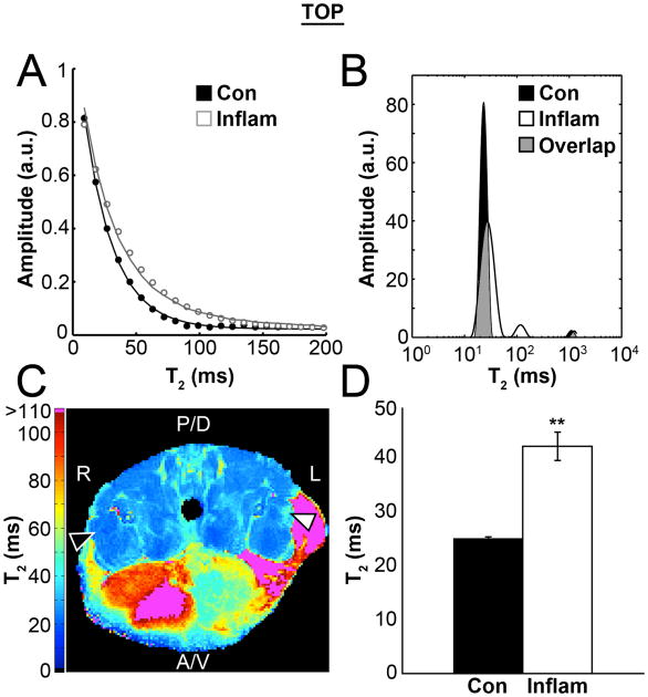
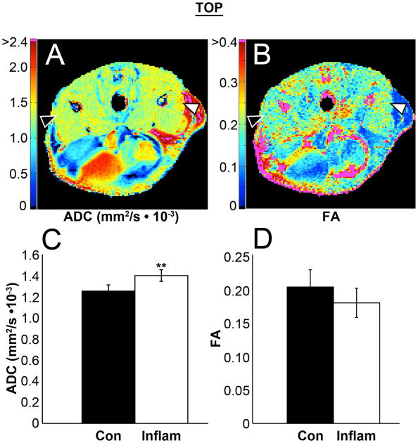
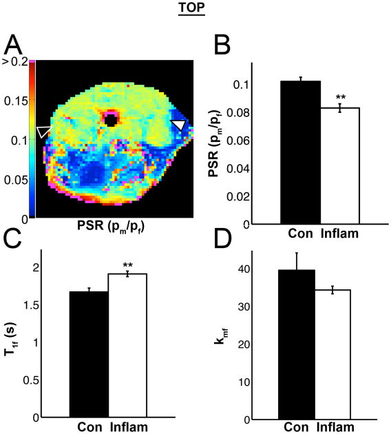
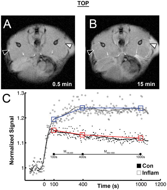
Myopathies often display a common set of complex pathologies that include muscle weakness, inflammation, compromised membrane integrity, fat deposition, and fibrosis. Multi-parametric, quantitative, non-invasive imaging approaches may be able to resolve these individual pathological components. The goal of this study was to use multi-parametric MRI to investigate inflammation as an isolated pathological feature. Proton relaxation, diffusion tensor imaging (DTI), quantitative magnetization transfer (qMT-MRI), and dynamic contrast enhanced (DCE-MRI) parameters were calculated from data acquired in a single imaging session conducted 6-8 hours following the injection of λ-carrageenan, a local inflammatory agent. T2 increased in the inflamed muscle and transitioned to bi-exponential behavior. In diffusion measurements, all three eigenvalues and the apparent diffusion coefficient increased, but λ3 had the largest relative change. Analysis of the qMT data revealed that the T1 of the free pool and the observed T1 both increased in the inflamed tissue, while the ratio of exchanging spins in the solid pool to those in the free water pool (the pool size ratio) significantly decreased. DCE-MRI data also supported observations of an increase in extracellular volume. These findings enriched the understanding of the relation between multiple quantitative MRI parameters and an isolated inflammatory pathology, and may potentially be employed for other single or complex myopathy models.
肌病通常表现出一组常见的复杂病理特征,包括肌肉无力、炎症、膜完整性受损、脂肪沉积和纤维化。多参数、定量、非侵入性成像方法或许能够分辨这些个体病理成分。本研究的目的是使用多参数磁共振成像(MRI)来研究炎症作为一种孤立的病理特征。质子弛豫、扩散张量成像(DTI)、定量磁化传递(qMT-MRI)和动态对比增强(DCE-MRI)参数是根据在注射局部炎症介质λ-角叉菜胶后6-8小时进行的单次成像扫描所采集的数据计算得出的。在发炎的肌肉中,T2增加并转变为双指数行为。在扩散测量中,所有三个特征值和表观扩散系数均增加,但λ3具有最大的相对变化。对qMT数据的分析显示,在发炎组织中,自由池的T1和观测到的T1均增加,而固体池中交换自旋与自由水池中交换自旋的比率(池大小比率)显著降低。DCE-MRI数据也支持细胞外体积增加的观察结果。这些发现丰富了对多个定量MRI参数与孤立炎症病理之间关系的理解,并且可能潜在地应用于其他单一或复杂的肌病模型。