• 文献检索
  • 文档翻译
  • 深度研究
  • 学术资讯
  • Suppr Zotero 插件Zotero 插件
  • 邀请有礼
  • 套餐&价格
  • 历史记录
应用&插件
Suppr Zotero 插件Zotero 插件浏览器插件Mac 客户端Windows 客户端微信小程序
定价
高级版会员购买积分包购买API积分包
服务
文献检索文档翻译深度研究API 文档MCP 服务
关于我们
关于 Suppr公司介绍联系我们用户协议隐私条款
关注我们

Suppr 超能文献

核心技术专利:CN118964589B侵权必究
粤ICP备2023148730 号-1Suppr @ 2026

文献检索

告别复杂PubMed语法,用中文像聊天一样搜索,搜遍4000万医学文献。AI智能推荐,让科研检索更轻松。

立即免费搜索

文件翻译

保留排版,准确专业,支持PDF/Word/PPT等文件格式,支持 12+语言互译。

免费翻译文档

深度研究

AI帮你快速写综述,25分钟生成高质量综述,智能提取关键信息,辅助科研写作。

立即免费体验

弥散张量成像用于表征肿瘤微结构并提高乳腺 MRI 的诊断性能:一项前瞻性观察研究。

Diffusion tensor imaging for characterizing tumor microstructure and improving diagnostic performance on breast MRI: a prospective observational study.

机构信息

Department of Radiology, University of Washington School of Medicine, 825 Eastlake Avenue East, Seattle, WA, 98109, USA.

Department of Pathology, University of Washington School of Medicine, 1959 NE Pacific St. Box 356100, Seattle, WA, 98195, USA.

出版信息

Breast Cancer Res. 2019 Sep 4;21(1):102. doi: 10.1186/s13058-019-1183-3.

DOI:10.1186/s13058-019-1183-3
PMID:31484577
原文链接:https://pmc.ncbi.nlm.nih.gov/articles/PMC6727336/
Abstract

BACKGROUND

Diffusion-weighted imaging (DWI) can increase breast MRI diagnostic specificity due to the tendency of malignancies to restrict diffusion. Diffusion tensor imaging (DTI) provides further information over conventional DWI regarding diffusion directionality and anisotropy. Our study evaluates DTI features of suspicious breast lesions detected on MRI to determine the added diagnostic value of DTI for breast imaging.

METHODS

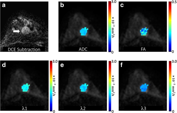
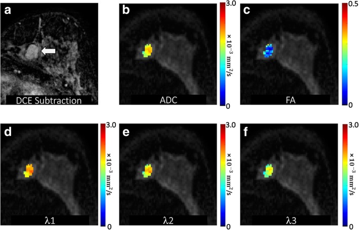
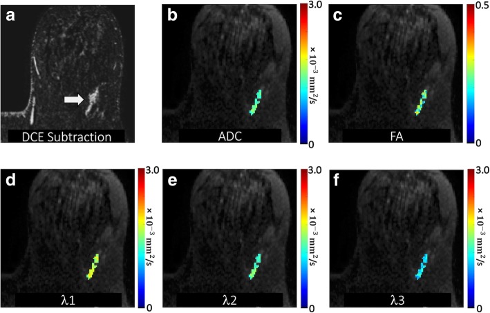
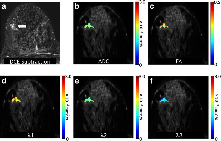
With IRB approval, we prospectively enrolled patients over a 3-year period who had suspicious (BI-RADS category 4 or 5) MRI-detected breast lesions with histopathological results. Patients underwent multiparametric 3 T MRI with dynamic contrast-enhanced (DCE) and DTI sequences. Clinical factors (age, menopausal status, breast density, clinical indication, background parenchymal enhancement) and DCE-MRI lesion parameters (size, type, presence of washout, BI-RADS category) were recorded prospectively by interpreting radiologists. DTI parameters (apparent diffusion coefficient [ADC], fractional anisotropy [FA], axial diffusivity [λ], radial diffusivity [(λ + λ)/2], and empirical difference [λ - λ]) were measured retrospectively. Generalized estimating equations (GEE) and least absolute shrinkage and selection operator (LASSO) methods were used for univariate and multivariate logistic regression, respectively. Diagnostic performance was internally validated using the area under the curve (AUC) with bootstrap adjustment.

RESULTS

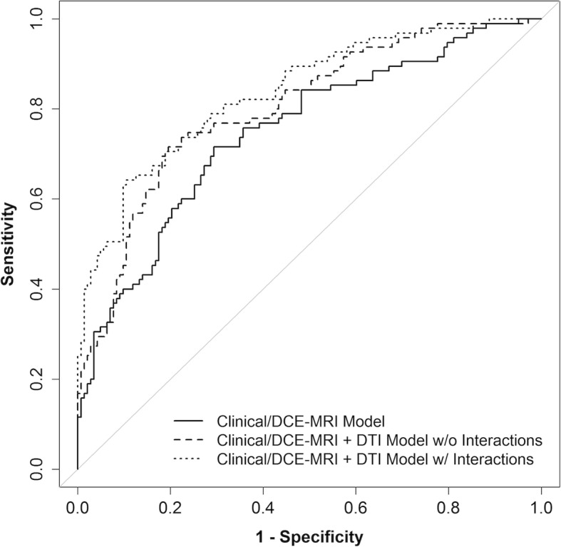
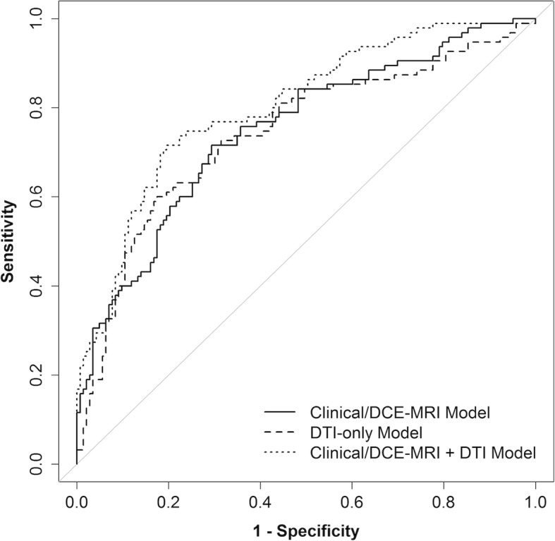
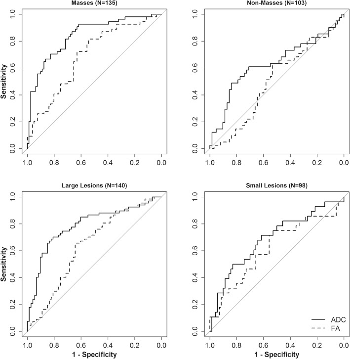
The study included 238 suspicious breast lesions (95 malignant, 143 benign) in 194 women. In univariate analysis, lower ADC, axial diffusivity, and radial diffusivity were associated with malignancy (OR = 0.37-0.42 per 1-SD increase, p < 0.001 for each), as was higher FA (OR = 1.45, p = 0.007). In multivariate analysis, LASSO selected only ADC (OR = 0.41) as a predictor for a DTI-only model, while both ADC (OR = 0.41) and FA (OR = 0.88) were selected for a model combining clinical and imaging parameters. Post-hoc analysis revealed varying association of FA with malignancy depending on the lesion type. The combined model (AUC = 0.81) had a significantly better performance than Clinical/DCE-MRI-only (AUC = 0.76, p < 0.001) and DTI-only (AUC = 0.75, p = 0.002) models.

CONCLUSIONS

DTI significantly improves diagnostic performance in multivariate modeling. ADC is the most important diffusion parameter for distinguishing benign and malignant breast lesions, while anisotropy measures may help further characterize tumor microstructure and microenvironment.

摘要

背景

扩散加权成像(DWI)可以通过限制恶性肿瘤的扩散来提高乳腺 MRI 的诊断特异性。扩散张量成像(DTI)提供了关于扩散方向和各向异性的常规 DWI 之外的更多信息。我们的研究评估了 MRI 检测到的可疑乳腺病变的 DTI 特征,以确定 DTI 对乳腺成像的附加诊断价值。

方法

在获得机构审查委员会批准后,我们前瞻性地招募了在 3 年期间具有可疑(BI-RADS 类别 4 或 5)MRI 检测到的乳腺病变且具有组织病理学结果的患者。患者接受了多参数 3T MRI 检查,包括动态对比增强(DCE)和 DTI 序列。由解释放射科医师前瞻性地记录临床因素(年龄、绝经状态、乳腺密度、临床指征、背景实质增强)和 DCE-MRI 病变参数(大小、类型、有无洗脱、BI-RADS 类别)。回顾性测量 DTI 参数(表观扩散系数[ADC]、各向异性分数[FA]、轴向扩散系数[λ]、径向扩散系数[(λ+λ)/2]和经验差[λ-λ])。使用广义估计方程(GEE)和最小绝对收缩和选择算子(LASSO)方法分别进行单变量和多变量逻辑回归。使用内部验证曲线下面积(AUC)进行调整后的 bootstrap 验证来评估诊断性能。

结果

该研究包括 194 名女性中的 238 个可疑乳腺病变(95 个恶性,143 个良性)。在单变量分析中,ADC、轴向扩散系数和径向扩散系数降低与恶性肿瘤相关(每增加 1-SD,OR=0.37-0.42,p<0.001),FA 升高(OR=1.45,p=0.007)也与恶性肿瘤相关。在多变量分析中,LASSO 仅选择 ADC(OR=0.41)作为 DTI 模型的预测因子,而 ADC(OR=0.41)和 FA(OR=0.88)都被选为结合临床和影像学参数的模型的预测因子。事后分析显示,FA 与恶性肿瘤的关联因病变类型而异。联合模型(AUC=0.81)的性能明显优于仅临床/DCE-MRI 模型(AUC=0.76,p<0.001)和仅 DTI 模型(AUC=0.75,p=0.002)。

结论

DTI 在多变量建模中显著提高了诊断性能。ADC 是区分良性和恶性乳腺病变的最重要的扩散参数,而各向异性测量值可能有助于进一步描述肿瘤的微观结构和微环境。

https://cdn.ncbi.nlm.nih.gov/pmc/blobs/1144/6727336/b6f6c4a6845e/13058_2019_1183_Fig7_HTML.jpg
https://cdn.ncbi.nlm.nih.gov/pmc/blobs/1144/6727336/5898def16681/13058_2019_1183_Fig1_HTML.jpg
https://cdn.ncbi.nlm.nih.gov/pmc/blobs/1144/6727336/98ab517d878f/13058_2019_1183_Fig2_HTML.jpg
https://cdn.ncbi.nlm.nih.gov/pmc/blobs/1144/6727336/2a074dca3361/13058_2019_1183_Fig3_HTML.jpg
https://cdn.ncbi.nlm.nih.gov/pmc/blobs/1144/6727336/8a80bcf1603f/13058_2019_1183_Fig4_HTML.jpg
https://cdn.ncbi.nlm.nih.gov/pmc/blobs/1144/6727336/4b0cae69b557/13058_2019_1183_Fig5_HTML.jpg
https://cdn.ncbi.nlm.nih.gov/pmc/blobs/1144/6727336/5c360258961d/13058_2019_1183_Fig6_HTML.jpg
https://cdn.ncbi.nlm.nih.gov/pmc/blobs/1144/6727336/b6f6c4a6845e/13058_2019_1183_Fig7_HTML.jpg

相似文献

1
Diffusion tensor imaging for characterizing tumor microstructure and improving diagnostic performance on breast MRI: a prospective observational study.弥散张量成像用于表征肿瘤微结构并提高乳腺 MRI 的诊断性能:一项前瞻性观察研究。
Breast Cancer Res. 2019 Sep 4;21(1):102. doi: 10.1186/s13058-019-1183-3.
2
Comparison of the diagnostic performances of diffusion parameters in diffusion weighted imaging and diffusion tensor imaging of breast lesions.扩散加权成像和扩散张量成像中扩散参数对乳腺病变诊断性能的比较。
Eur J Radiol. 2013 Dec;82(12):e801-6. doi: 10.1016/j.ejrad.2013.09.001. Epub 2013 Sep 13.
3
Breast MRI during lactation: effects on tumor conspicuity using dynamic contrast-enhanced (DCE) in comparison with diffusion tensor imaging (DTI) parametric maps.哺乳期乳腺 MRI:动态对比增强(DCE)与扩散张量成像(DTI)参数图对肿瘤显示效果的比较。
Eur Radiol. 2020 Feb;30(2):767-777. doi: 10.1007/s00330-019-06435-x. Epub 2019 Sep 16.
4
Diagnostic performance of quantitative diffusion tensor imaging for the differentiation of breast lesions at 3 T MRI.3T磁共振成像定量扩散张量成像对乳腺病变鉴别诊断的效能
Clin Imaging. 2019 Jan-Feb;53:25-31. doi: 10.1016/j.clinimag.2018.10.002. Epub 2018 Oct 3.
5
Diagnostic performance of diffusion tensor imaging parameters in breast cancer and correlation with the prognostic factors.扩散张量成像参数在乳腺癌中的诊断效能及其与预后因素的相关性
J Magn Reson Imaging. 2017 Mar;45(3):660-672. doi: 10.1002/jmri.25481. Epub 2016 Sep 23.
6
[3.0 T MR diffusion tentor imaging in the differential diagnosis of breast mass lesions].[3.0T磁共振扩散张量成像在乳腺肿块病变鉴别诊断中的应用]
Zhonghua Yi Xue Za Zhi. 2016 May 24;96(19):1510-4. doi: 10.3760/cma.j.issn.0376-2491.2016.19.010.
7
Apparent diffusion coefficient values may help predict which MRI-detected high-risk breast lesions will upgrade at surgical excision.表观扩散系数值可能有助于预测哪些 MRI 检测到的高风险乳腺病变在手术切除时会升级。
J Magn Reson Imaging. 2017 Oct;46(4):1028-1036. doi: 10.1002/jmri.25656. Epub 2017 Feb 9.
8
Diffusion-weighted imaging versus dynamic contrast-enhanced MRI: a new horizon for characterisation of suspicious breast lesions.扩散加权成像与动态对比增强 MRI:可疑乳腺病变特征分析的新视野。
Clin Radiol. 2021 Jan;76(1):80.e1-80.e8. doi: 10.1016/j.crad.2020.08.031. Epub 2020 Oct 17.
9
The Role of Combined Diffusion-Weighted Imaging and Dynamic Contrast-Enhanced MRI for Differentiating Malignant From Benign Breast Lesions Presenting Washout Curve.联合扩散加权成像和动态对比增强 MRI 在鉴别呈洗脱曲线的良恶性乳腺病变中的作用。
Can Assoc Radiol J. 2021 Aug;72(3):460-469. doi: 10.1177/0846537120907098. Epub 2020 Mar 11.
10
Diffusion weighted imaging for the differentiation of breast tumors: From apparent diffusion coefficient to high order diffusion tensor imaging.用于乳腺肿瘤鉴别诊断的扩散加权成像:从表观扩散系数到高阶扩散张量成像
J Magn Reson Imaging. 2016 May;43(5):1111-21. doi: 10.1002/jmri.25067. Epub 2015 Oct 22.

引用本文的文献

1
Establishment of an interpretable MRI radiomics-based machine learning model capable of predicting axillary lymph node metastasis in invasive breast cancer.建立一种基于可解释性磁共振成像放射组学的机器学习模型,该模型能够预测浸润性乳腺癌腋窝淋巴结转移。
Sci Rep. 2025 Jul 18;15(1):26030. doi: 10.1038/s41598-025-10818-0.
2
Mean Apparent Propagator MRI: Quantitative Assessment of Tumor-Stroma Ratio in Invasive Ductal Breast Carcinoma.平均表观弥散系数 MRI:浸润性导管乳腺癌肿瘤-基质比的定量评估。
Radiol Imaging Cancer. 2024 Jul;6(4):e230165. doi: 10.1148/rycan.230165.
3
Diffusion tensor imaging: survival analysis prediction in breast cancer patients.

本文引用的文献

1
Utility of Diffusion-weighted Imaging to Decrease Unnecessary Biopsies Prompted by Breast MRI: A Trial of the ECOG-ACRIN Cancer Research Group (A6702).扩散加权成像在减少乳腺 MRI 引导的不必要活检中的应用:ECOG-ACRIN 癌症研究组(A6702)的一项试验。
Clin Cancer Res. 2019 Mar 15;25(6):1756-1765. doi: 10.1158/1078-0432.CCR-18-2967. Epub 2019 Jan 15.
2
Test-retest repeatability and reproducibility of ADC measures by breast DWI: Results from the ACRIN 6698 trial.乳腺 DWI 中 ADC 测量的重测重复性和再现性:来自 ACRIN 6698 试验的结果。
J Magn Reson Imaging. 2019 Jun;49(6):1617-1628. doi: 10.1002/jmri.26539. Epub 2018 Oct 22.
3
弥散张量成像:乳腺癌患者的生存分析预测。
Radiologie (Heidelb). 2024 Nov;64(Suppl 1):54-59. doi: 10.1007/s00117-023-01254-0. Epub 2024 Jan 26.
4
Multiparametric Approach to Breast Cancer With Emphasis on Magnetic Resonance Imaging in the Era of Personalized Breast Cancer Treatment.多参数方法在乳腺癌诊治中的应用——个体化乳腺癌治疗时代的磁共振成像重点
Invest Radiol. 2024 Jan 1;59(1):26-37. doi: 10.1097/RLI.0000000000001044. Epub 2023 Nov 22.
5
Head-to-head comparison of perfluorobutane contrast-enhanced US and multiparametric MRI for breast cancer: a prospective, multicenter study.对比氟丙烷对比增强超声与多参数 MRI 用于乳腺癌的头对头研究:一项前瞻性、多中心研究。
Breast Cancer Res. 2023 May 30;25(1):61. doi: 10.1186/s13058-023-01650-3.
6
The road to breast cancer screening with diffusion MRI.通过扩散磁共振成像进行乳腺癌筛查之路。
Front Oncol. 2023 Feb 21;13:993540. doi: 10.3389/fonc.2023.993540. eCollection 2023.
7
CEST-MRI for body oncologic imaging: are we there yet?体部肿瘤磁共振成像中的 CEST-MRI:我们成功了吗?
NMR Biomed. 2023 Jun;36(6):e4906. doi: 10.1002/nbm.4906. Epub 2023 Feb 15.
8
Breast MRI: an illustration of benign findings.乳腺磁共振成像:良性表现举例。
Br J Radiol. 2023 Feb;96(1142):20220280. doi: 10.1259/bjr.20220280. Epub 2022 Dec 9.
9
Contrast-Enhanced Mammography versus Breast Magnetic Resonance Imaging: A Systematic Review and Meta-Analysis.对比增强乳腺钼靶摄影与乳腺磁共振成像:一项系统评价与荟萃分析
Diagnostics (Basel). 2022 Aug 4;12(8):1890. doi: 10.3390/diagnostics12081890.
10
The Impact of Dense Breasts on the Stage of Breast Cancer at Diagnosis: A Review and Options for Supplemental Screening.致密型乳腺对乳腺癌诊断分期的影响:综述及补充筛查的选择。
Curr Oncol. 2022 May 17;29(5):3595-3636. doi: 10.3390/curroncol29050291.
Diffusion-weighted MRI Findings Predict Pathologic Response in Neoadjuvant Treatment of Breast Cancer: The ACRIN 6698 Multicenter Trial.
扩散加权 MRI 表现预测乳腺癌新辅助治疗的病理反应:ACRIN 6698 多中心试验。
Radiology. 2018 Dec;289(3):618-627. doi: 10.1148/radiol.2018180273. Epub 2018 Sep 4.
4
Effect of Background Parenchymal Enhancement on Breast MR Imaging Interpretive Performance in Community-based Practices.背景实质强化对社区实践中乳腺磁共振成像解读性能的影响。
Radiology. 2018 Mar;286(3):822-829. doi: 10.1148/radiol.2017170811. Epub 2017 Oct 25.
5
Performance Benchmarks for Screening Breast MR Imaging in Community Practice.社区实践中乳腺磁共振成像筛查的性能基准
Radiology. 2017 Oct;285(1):44-52. doi: 10.1148/radiol.2017162033. Epub 2017 Jun 5.
6
Screening Breast MRI Outcomes in Routine Clinical Practice: Comparison to BI-RADS Benchmarks.常规临床实践中乳腺MRI筛查结果:与BI-RADS标准的比较。
Acad Radiol. 2017 Apr;24(4):411-417. doi: 10.1016/j.acra.2016.10.014. Epub 2016 Dec 13.
7
Diffusion-weighted breast MRI: Clinical applications and emerging techniques.扩散加权乳腺磁共振成像:临床应用与新兴技术。
J Magn Reson Imaging. 2017 Feb;45(2):337-355. doi: 10.1002/jmri.25479. Epub 2016 Sep 30.
8
Distortion correction in diffusion-weighted imaging of the breast: Performance assessment of prospective, retrospective, and combined (prospective + retrospective) approaches.乳腺扩散加权成像中的畸变校正:前瞻性、回顾性及联合(前瞻性+回顾性)方法的性能评估
Magn Reson Med. 2017 Jul;78(1):247-253. doi: 10.1002/mrm.26328. Epub 2016 Jul 12.
9
Diffusion tensor imaging of breast lesions: evaluation of apparent diffusion coefficient and fractional anisotropy and tissue cellularity.乳腺病变的扩散张量成像:表观扩散系数、各向异性分数及组织细胞密度的评估
Br J Radiol. 2016 Aug;89(1064):20160076. doi: 10.1259/bjr.20160076. Epub 2016 Jun 15.
10
Diffusion-Weighted Breast Magnetic Resonance Imaging: A Semiautomated Voxel Selection Technique Improves Interreader Reproducibility of Apparent Diffusion Coefficient Measurements.扩散加权乳腺磁共振成像:一种半自动体素选择技术可提高表观扩散系数测量的阅片者间可重复性。
J Comput Assist Tomogr. 2016 May-Jun;40(3):428-35. doi: 10.1097/RCT.0000000000000372.