Wang Jiang-Feng, Feng Jian-Guo, Han Jing, Zhang Bei-Bei, Mao Wei-Min
Cancer Research Institute, Zhejiang Cancer Hospital, No. 38 Guangji Road, Hangzhou, Zhejiang 310022, China ; Key Laboratory Diagnosis and Treatment Technology on Thoracic Oncology, Hangzhou, Zhejiang 310022, China.
Biomed Res Int. 2014;2014:582730. doi: 10.1155/2014/582730. Epub 2014 Apr 15.
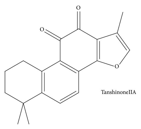
To explore the possible mechanisms of Tanshinone IIA (TanIIA) on esophageal carcinoma cell lines.
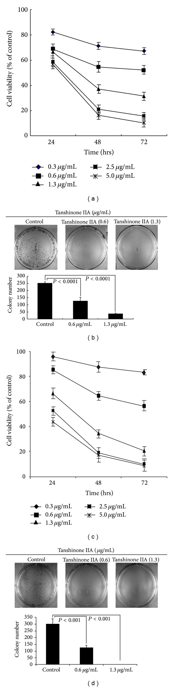
Two human esophageal carcinoma cell lines (EC-1 cells and ECa-109 cells) were treated with different concentrations of TanIIA. Cell proliferation was measured by CCK-8, colony-forming efficiency was calculated, cell cycle and apoptosis were measured, and changes in cell cycle- and apoptosis-related gene expression were measured by Western blotting.
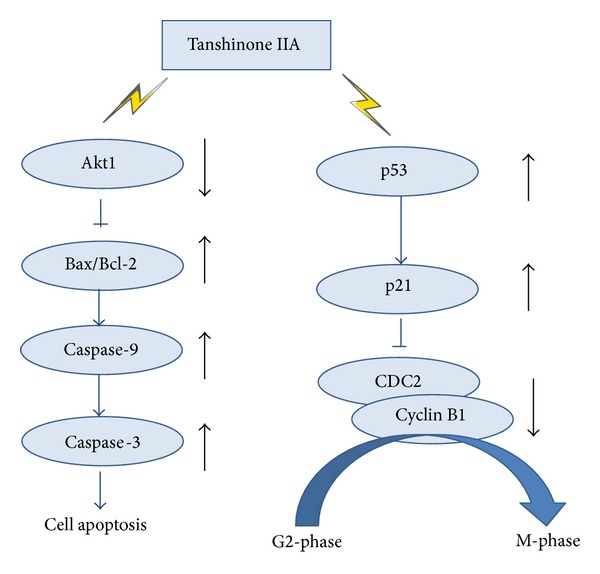
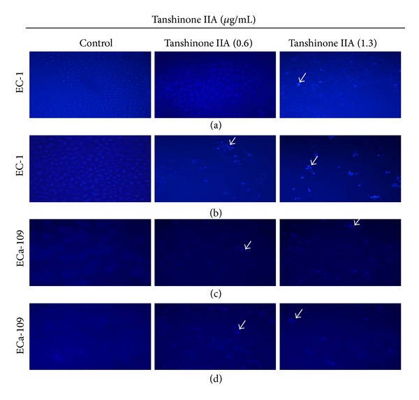
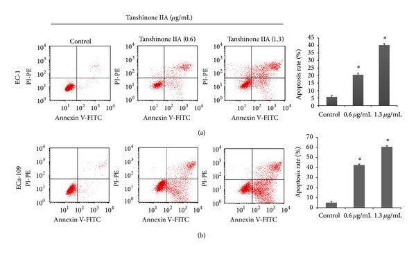
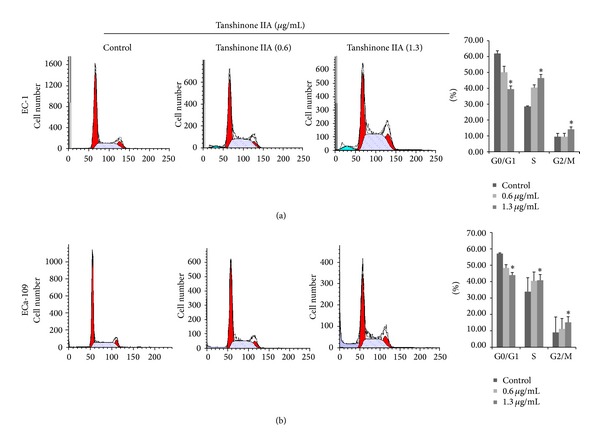
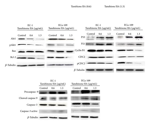
The CCK-8 and colony formation assay indicated that TanIIA inhibited the cell proliferation of human esophageal cancer cells (IC50 below 1 μg/mL) at 48 h. Hoechst 33258 and flow cytometry showed that TanIIA induced apoptosis in both esophageal cancer cell lines. Flow cytometry showed that TanIIA arrested cell cycle in S phase and G2/M phase. Western blotting analysis showed that Akt1 and its phosphorylation were inhibited, the Bax/Bcl-2 ratio increased, and both caspase-9 and caspase-3 were activated after treatment with 1.3 μg/mL TanIIA at 48 h. Meanwhile, p53 and p21 protein levels increased, whereas cyclin B1, CDC2, and CDC2 phosphorylation were inhibited.
TanIIA inhibits the growth of esophageal cancer cells and induces apoptosis in a time-dependent and concentration-dependent manner, possibly by affecting cell cycle- and apoptosis-related signaling pathways.
探讨丹参酮IIA(TanIIA)对食管癌细胞系的可能作用机制。
用不同浓度的TanIIA处理两种人食管癌细胞系(EC-1细胞和ECa-109细胞)。采用CCK-8法检测细胞增殖,计算集落形成效率,检测细胞周期和凋亡情况,并通过蛋白质免疫印迹法检测细胞周期和凋亡相关基因表达的变化。
CCK-8和集落形成实验表明,TanIIA在48小时时抑制人食管癌细胞的增殖(IC50低于1μg/mL)。Hoechst 33258染色和流式细胞术显示,TanIIA诱导两种食管癌细胞系凋亡。流式细胞术显示,TanIIA使细胞周期停滞在S期和G2/M期。蛋白质免疫印迹分析显示,用1.3μg/mL TanIIA处理48小时后,Akt1及其磷酸化水平受到抑制,Bax/Bcl-2比值增加,caspase-9和caspase-3均被激活。同时,p53和p21蛋白水平升高,而细胞周期蛋白B1、细胞周期蛋白依赖性激酶2(CDC2)及其磷酸化水平受到抑制。
TanIIA以时间和浓度依赖性方式抑制食管癌细胞生长并诱导凋亡,可能是通过影响细胞周期和凋亡相关信号通路实现的。