Miyoshi Ko, Kasahara Kyosuke, Murakami Shinki, Takeshima Mika, Kumamoto Natsuko, Sato Asako, Miyazaki Ikuko, Matsuzaki Shinsuke, Sasaoka Toshikuni, Katayama Taiichi, Asanuma Masato
Department of Child Development and Molecular Brain Science, United Graduate School of Child Development, Osaka University, Suita, Japan; Molecular Research Center for Children's Mental Development, United Graduate School of Child Development, Osaka University, Suita, Japan; Department of Brain Science, Okayama University Graduate School of Medicine, Dentistry and Pharmaceutical Sciences, Okayama, Japan.
Department of Brain Science, Okayama University Graduate School of Medicine, Dentistry and Pharmaceutical Sciences, Okayama, Japan.
PLoS One. 2014 May 15;9(5):e97918. doi: 10.1371/journal.pone.0097918. eCollection 2014.
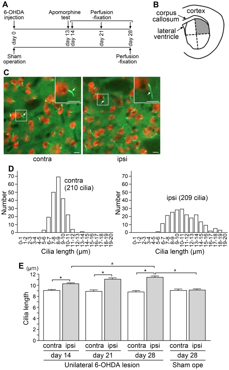
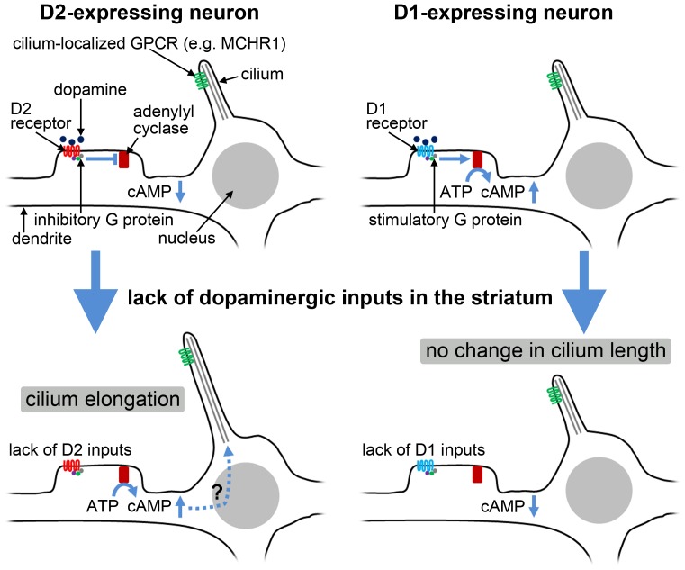
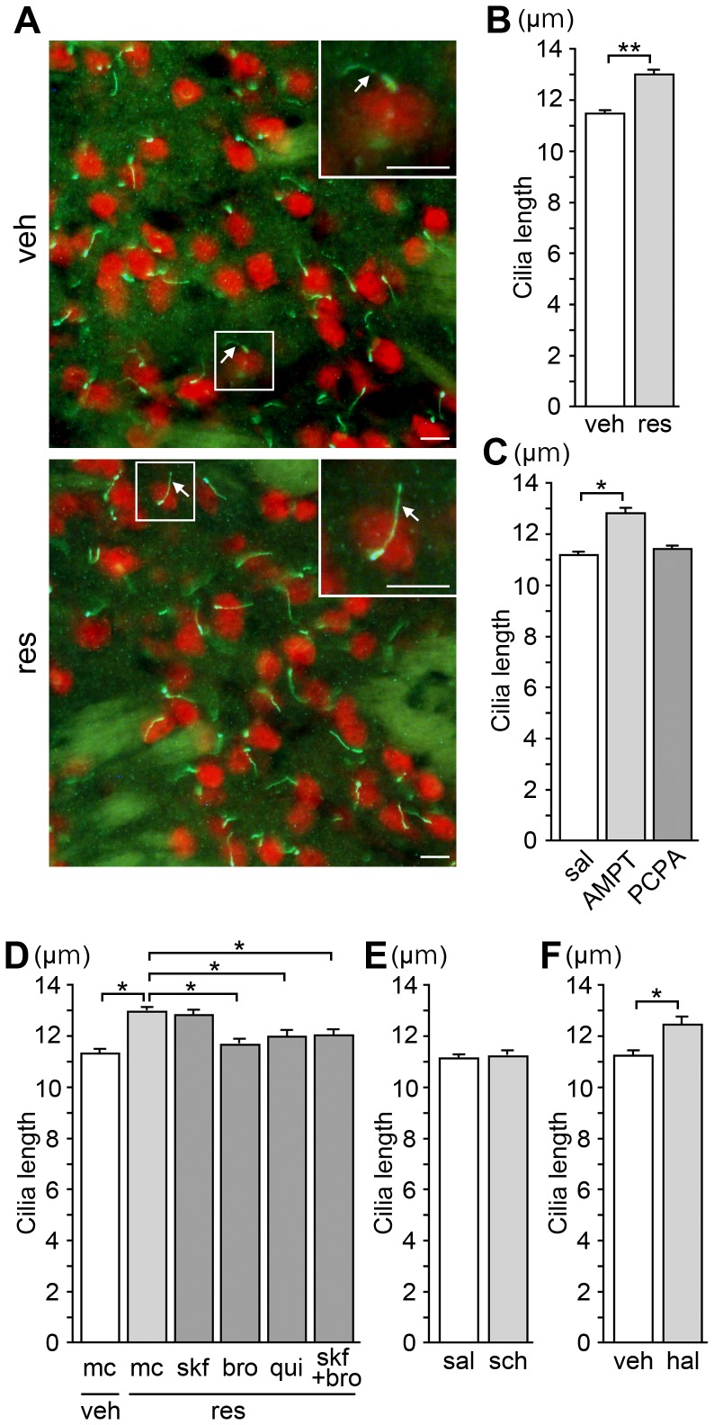
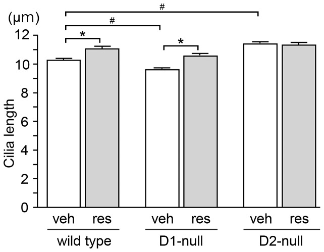
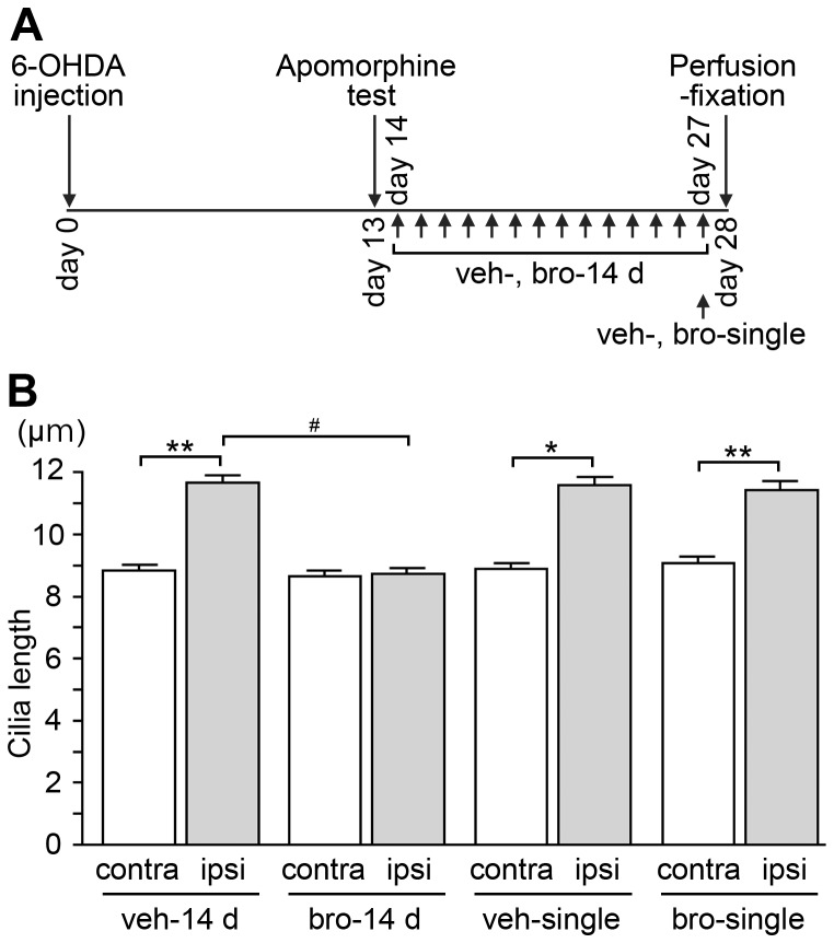
In the rodent brain, certain G protein-coupled receptors and adenylyl cyclase type 3 are known to localize to the neuronal primary cilium, a primitive sensory organelle protruding singly from almost all neurons. A recent chemical screening study demonstrated that many compounds targeting dopamine receptors regulate the assembly of Chlamydomonas reinhardtii flagella, structures which are analogous to vertebrate cilia. Here we investigated the effects of dopaminergic inputs loss on the architecture of neuronal primary cilia in the rodent striatum, a brain region that receives major dopaminergic projections from the midbrain. We first analyzed the lengths of neuronal cilia in the dorsolateral striatum of hemi-parkinsonian rats with unilateral lesions of the nigrostriatal dopamine pathway. In these rats, the striatal neuronal cilia were significantly longer on the lesioned side than on the non-lesioned side. In mice, the repeated injection of reserpine, a dopamine-depleting agent, elongated neuronal cilia in the striatum. The combined administration of agonists for dopamine receptor type 2 (D2) with reserpine attenuated the elongation of striatal neuronal cilia. Repeated treatment with an antagonist of D2, but not of dopamine receptor type 1 (D1), elongated the striatal neuronal cilia. In addition, D2-null mice displayed longer neuronal cilia in the striatum compared to wild-type controls. Reserpine treatment elongated the striatal neuronal cilia in D1-null mice but not in D2-null mice. Repeated treatment with a D2 agonist suppressed the elongation of striatal neuronal cilia on the lesioned side of hemi-parkinsonian rats. These results suggest that the elongation of striatal neuronal cilia following the lack of dopaminergic inputs is attributable to the absence of dopaminergic transmission via D2 receptors. Our results provide the first evidence that the length of neuronal cilia can be modified by the lack of a neurotransmitter's input.
在啮齿动物大脑中,某些G蛋白偶联受体和3型腺苷酸环化酶已知定位于神经元的初级纤毛,这是一种几乎从所有神经元单独伸出的原始感觉细胞器。最近的一项化学筛选研究表明,许多靶向多巴胺受体的化合物可调节莱茵衣藻鞭毛的组装,而鞭毛结构类似于脊椎动物的纤毛。在这里,我们研究了多巴胺能输入缺失对啮齿动物纹状体中神经元初级纤毛结构的影响,纹状体是一个从中脑接收主要多巴胺能投射的脑区。我们首先分析了黑质纹状体多巴胺通路单侧损伤的半帕金森病大鼠背外侧纹状体中神经元纤毛的长度。在这些大鼠中,损伤侧的纹状体神经元纤毛明显比未损伤侧长。在小鼠中,重复注射利血平(一种多巴胺耗竭剂)可使纹状体中的神经元纤毛伸长。多巴胺2型受体(D2)激动剂与利血平联合给药可减弱纹状体神经元纤毛的伸长。用D2拮抗剂而非多巴胺1型受体(D1)拮抗剂重复治疗可使纹状体神经元纤毛伸长。此外,与野生型对照相比,D2基因敲除小鼠的纹状体中神经元纤毛更长。利血平治疗可使D1基因敲除小鼠的纹状体神经元纤毛伸长,但对D2基因敲除小鼠无效。用D2激动剂重复治疗可抑制半帕金森病大鼠损伤侧纹状体神经元纤毛的伸长。这些结果表明,多巴胺能输入缺乏后纹状体神经元纤毛的伸长归因于通过D2受体的多巴胺能传递缺失。我们的结果提供了首个证据,即神经元纤毛的长度可因神经递质输入的缺乏而改变。