Molecular Biophysics Laboratory, Photonics Center, and Department of Physics, Boston University , Boston, Massachusetts 02215, United States.
Biochemistry. 2014 Jun 24;53(24):3961-70. doi: 10.1021/bi500445c. Epub 2014 Jun 16.
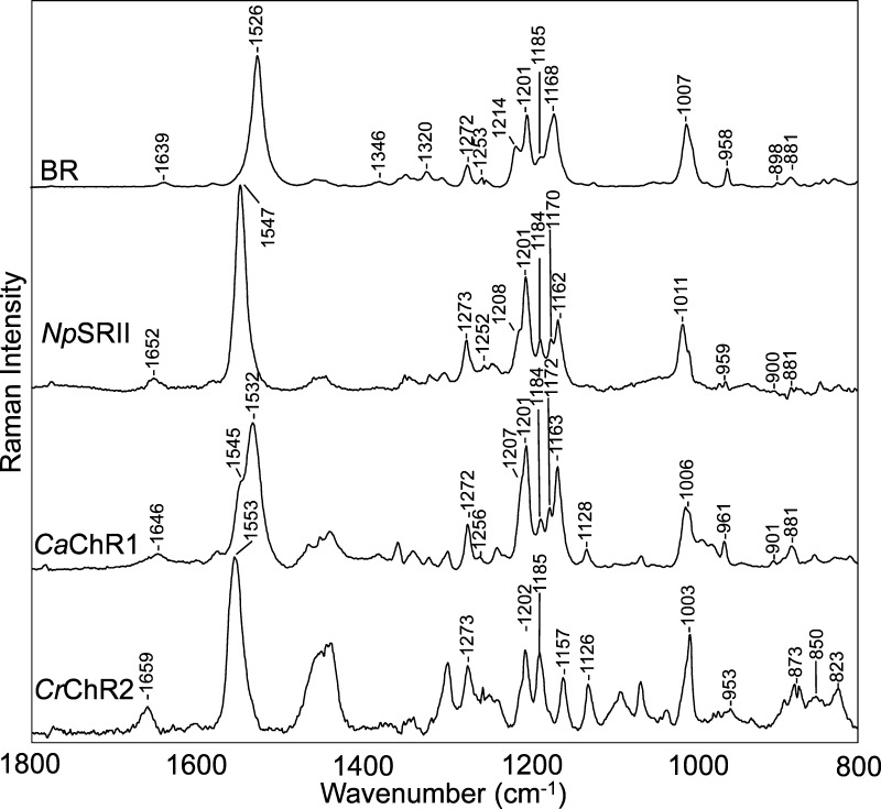
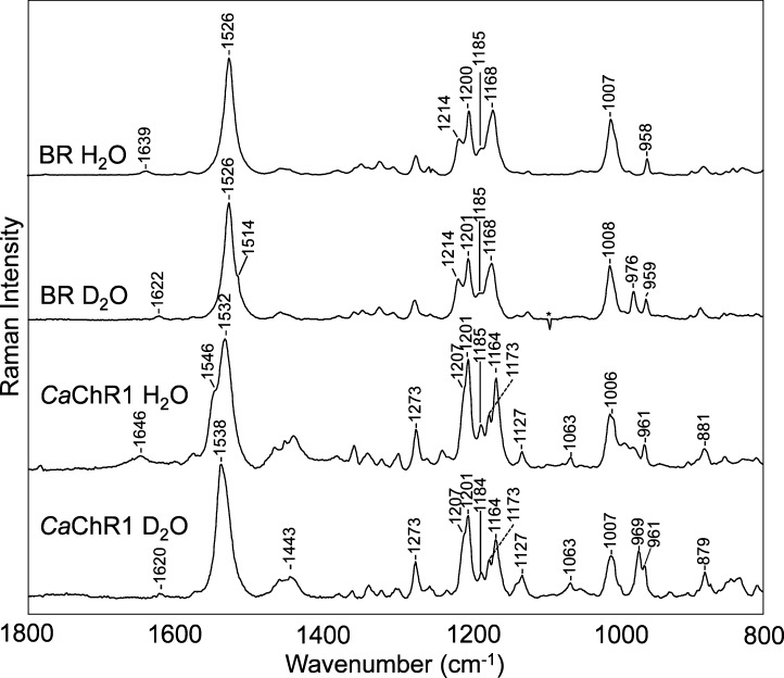
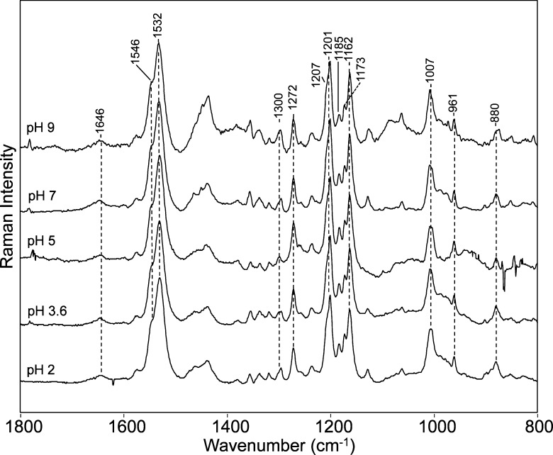
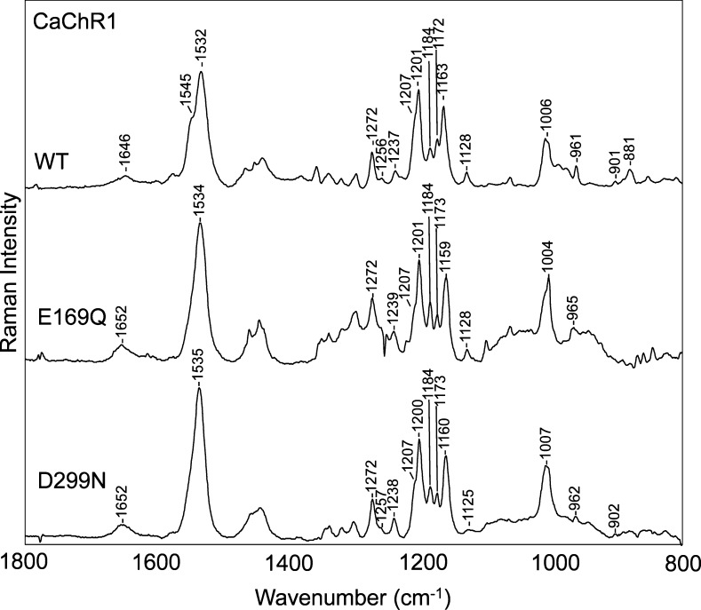
Channelrhodopsins (ChRs), which form a distinct branch of the microbial rhodopsin family, control phototaxis in green algae. Because ChRs can be expressed and function in neuronal membranes as light-gated cation channels, they have rapidly become an important optogenetic tool in neurobiology. While channelrhodopsin-2 from the unicellular alga Chlamydomonas reinhardtii (CrChR2) is the most commonly used and extensively studied optogenetic ChR, little is known about the properties of the diverse group of other ChRs. In this study, near-infrared confocal resonance Raman spectroscopy along with hydrogen-deuterium exchange and site-directed mutagenesis were used to study the structure of red-shifted ChR1 from Chlamydomonas augustae (CaChR1). These measurements reveal that (i) CaChR1 has an all-trans-retinal structure similar to those of the light-driven proton pump bacteriorhodopsin (BR) and sensory rhodopsin II but different from that of the mixed retinal composition of CrChR2, (ii) lowering the pH from 7 to 2 or substituting neutral residues for Glu169 or Asp299 does not significantly shift the ethylenic stretch frequency more than 1-2 cm(-1) in contrast to BR in which a downshift of 7-9 cm(-1) occurs reflecting neutralization of the Asp85 counterion, and (iii) the CaChR1 protonated Schiff base (SB) has stronger hydrogen bonding than BR. A model is proposed to explain these results whereby at pH 7 the predominant counterion to the SB is Asp299 (the homologue to Asp212 in BR) while Glu169 (the homologue to Asp85 in BR) exists in a neutral state. We observe an unusual constancy of the resonance Raman spectra over the broad range from pH 9 to 2 and discuss its implications. These results are in accord with recent visible absorption and current measurements of CaChR1 [Sineshchekov, O. A., et al. (2013) Intramolecular proton transfer in channelrhodopsins. Biophys. J. 104, 807-817; Li, H., et al. (2014) Role of a helix B lysine residue in the photoactive site in channelrhodopsins. Biophys. J. 106, 1607-1617].
通道蛋白视紫红质(ChRs),构成微生物视紫红质家族的一个独特分支,控制着绿藻的趋光性。由于 ChRs 可以在神经元膜中作为光门控阳离子通道表达和发挥作用,它们已迅速成为神经生物学中一种重要的光遗传学工具。虽然来自单细胞绿藻莱茵衣藻(Chlamydomonas reinhardtii)的通道蛋白视紫红质-2(CrChR2)是最常用和广泛研究的光遗传学 ChR,但对其他 ChR 多样化群体的特性知之甚少。在这项研究中,使用近红外共焦共振拉曼光谱以及氘氢交换和定点突变来研究来自 Augustae 衣藻(Chlamydomonas augustae)的红移型 ChR1(CaChR1)的结构。这些测量结果表明:(i)CaChR1 具有与光驱动质子泵菌视紫红质(BR)和感觉视紫红质 II 相似的全反式视黄醛结构,但与 CrChR2 中混合视黄醛组成不同,(ii)与 BR 中发生 7-9 cm-1 向下位移反映出 Asp85 抗衡离子的中和相比,将 pH 值从 7 降低到 2 或用中性残基替代 Glu169 或 Asp299 不会使乙烯基伸缩频率显著移动超过 1-2 cm-1,(iii)CaChR1 的质子化席夫碱(SB)具有比 BR 更强的氢键。提出了一个模型来解释这些结果,即在 pH 7 时,SB 的主要抗衡离子是 Asp299(与 BR 中的 Asp212 同源),而 Glu169(与 BR 中的 Asp85 同源)处于中性状态。我们观察到从 pH 9 到 2 的宽范围内共振拉曼光谱的异常恒定,并讨论了其意义。这些结果与最近的 CaChR1 可见吸收和电流测量结果一致[O. A. Sineshchekov 等人(2013 年)通道蛋白视紫红质中的分子内质子转移。生物物理 J. 104, 807-817;H. Li 等人(2014 年)通道蛋白视紫红质中光活性位点中螺旋 B 赖氨酸残基的作用。生物物理 J. 106, 1607-1617]。