Hansmann Jan, Michaely Henrik J, Morelli John N, Luckscheiter André, Schoenberg Stefan O, Attenberger Ulrike I
Institute of Clinical Radiology and Nuclear Medicine, University Medical Center Mannheim, Medical Faculty Mannheim - Heidelberg University, Mannheim, Germany.
The Russel H. Morgan Department of Radiology and Radiological Science, The Johns Hopkins Hospital, Baltimore, Maryland, United States of America.
PLoS One. 2014 Jun 3;9(6):e99079. doi: 10.1371/journal.pone.0099079. eCollection 2014.
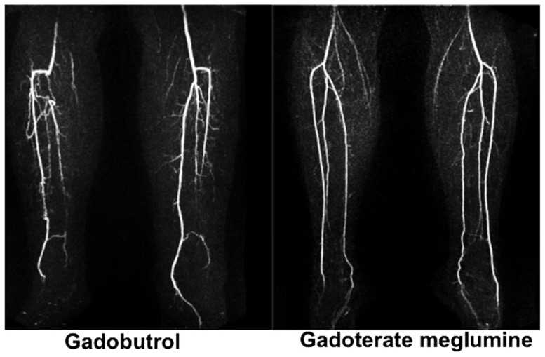
To compare enhancement characteristics and image quality of two macrocyclic gadolinium chelates, gadoterate meglumine and gadobutrol, in low-dose, time-resolved MRA of the calf station.
100 consecutive patients with peripheral arterial disease (stages II-IV) were retrospectively analysed. Fifty patients were included in each group - 32 men and 18 women for gadobutrol (mean age 67 years) and 34 men, 16 women for gadoterate meglumine (mean age 64 years). 0.03 mmol/kg bw of either gadobutrol or gadoterate meglumine was injected. Gadobutrol was diluted 1 ∶ 1 with normal saline (0.9% NaCl) to provide similar injection volume and bolus geometry compared to the undiluted 0.5 M dose of gadoterate meglumine. Signal-to-noise-ratio (SNR), contrast-to-noise-ratio (CNR) and image quality were analysed and compared between the two groups.
Mean SNR ranged from 83.0 ± 46.7 (peroneal artery) to 96.4 ± 64.5 (anterior tibial artery) for gadobutrol, and from 37.6 ± 13.8 (peroneal artery) to 45.3 ± 16.4 (anterior tibial artery) for the gadoterate meglumine group (p<0.0001). CNR values ranged from 30.1 ± 20.1 (peroneal artery) to 37.6 ± 26.0 (anterior tibial artery) for gadobutrol and from 14.9 ± 8.0 (peroneal artery) to 18.6 ± 16.4 (anterior tibial artery) for gadoterate meglumine (p<0.0001). No significant difference in image quality was found except for the peroneal arteries (p = 0.006 and p = 0.04). Interreader agreement was excellent (kappa 0.87-0.93).
The significantly better enhancement as assessed by SNR and CNR provided by gadobutrol compared to gadoterate meglumine does not translate into substantial differences in image quality in an equimolar, low-dose, time-resolved MRA protocol of the calves.
比较两种大环钆螯合物钆喷酸葡胺和钆布醇在小腿部位低剂量、时间分辨磁共振血管造影(MRA)中的强化特征及图像质量。
回顾性分析100例连续的外周动脉疾病(II-IV期)患者。每组纳入50例患者——钆布醇组32例男性和18例女性(平均年龄67岁),钆喷酸葡胺组34例男性和16例女性(平均年龄64岁)。分别注射0.03 mmol/kg体重的钆布醇或钆喷酸葡胺。钆布醇用生理盐水(0.9%氯化钠)1∶1稀释,以使其与未稀释的0.5 M剂量钆喷酸葡胺相比具有相似的注射体积和团注形态。分析并比较两组之间的信噪比(SNR)、对比噪声比(CNR)和图像质量。
钆布醇组的平均SNR范围为腓动脉83.0±46.7至胫前动脉96.4±64.5,钆喷酸葡胺组为腓动脉37.6±13.8至胫前动脉45.3±16.4(p<0.0001)。钆布醇组的CNR值范围为腓动脉30.1±20.1至胫前动脉37.6±26.0,钆喷酸葡胺组为腓动脉14.9±8.0至胫前动脉18.6±16.4(p<0.0001)。除腓动脉外,两组图像质量无显著差异(p = 0.006和p = 0.04)。阅片者间一致性良好(kappa值0.87 - 0.93)。
在小腿等摩尔、低剂量、时间分辨MRA方案中,与钆喷酸葡胺相比,钆布醇经SNR和CNR评估的强化效果明显更好,但这并未转化为图像质量的实质性差异。