Myers Anna Cardiakidis, Ghosh Fredrik, Andréasson Sten, Ponjavic Vesna
Department of Ophthalmology, Lund University , Lund , Sweden.
Curr Eye Res. 2014 Nov;39(11):1106-16. doi: 10.3109/02713683.2014.898309. Epub 2014 Jun 4.
To study the effects of the tumor necrosis factor alpha inhibitor adalimumab on rabbit retina after injection into the vitreous body.
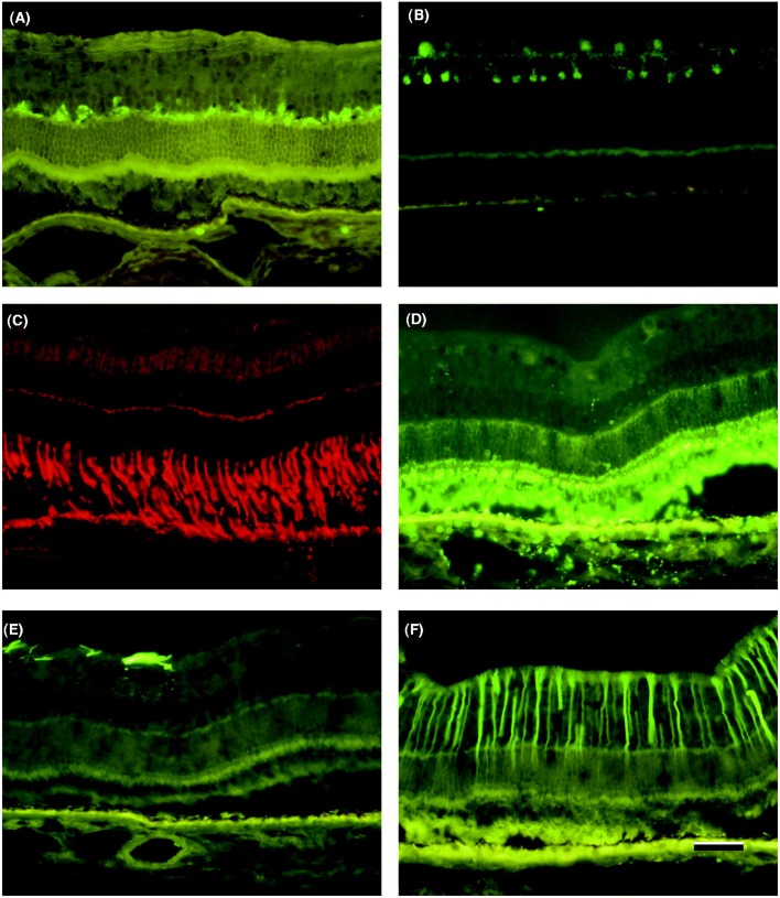
Forty-eight rabbits of mixed strain (9-12 months old, weighing ≈ 3.5 kg) were randomized into four groups. Adalimumab was injected at one of two concentrations (1.25 mg or 2.5 mg) into the eyes of two groups, and balanced salt solution into the eyes of the third group. The fourth group acted as controls. Full-field electroretinography (ffERG) was performed before injection and 1 and 6 weeks post-injection. At 6 weeks post-injection the rabbits were euthanized and the sectioned retinas were studied. Retinal histology was studied with hematoxylin-eosin staining. Immunohistochemical analysis was performed on rods, cones, rod bipolar cells, horizontal cells, amacrine cells and Müller cells.
No significant difference in ffERG amplitudes or implicit times was observed between the four groups at any time point. Histological and immunohistochemical findings were similar in all groups.
Injection of adalimumab into the vitreous body of healthy rabbits, at doses up to 2.5 mg, does not appear to be toxic to the rabbit retina.
研究肿瘤坏死因子α抑制剂阿达木单抗玻璃体腔内注射对兔视网膜的影响。
将48只混合品种兔(9 - 12月龄,体重约3.5 kg)随机分为四组。两组兔眼分别注射两种浓度(1.25 mg或2.5 mg)的阿达木单抗,第三组兔眼注射平衡盐溶液,第四组作为对照组。在注射前以及注射后1周和6周进行全视野视网膜电图(ffERG)检查。注射后6周处死兔子,对视网膜切片进行研究。用苏木精-伊红染色研究视网膜组织学。对视杆细胞、视锥细胞、视杆双极细胞、水平细胞、无长突细胞和Müller细胞进行免疫组织化学分析。
在任何时间点,四组之间的ffERG振幅或潜伏时间均未观察到显著差异。所有组的组织学和免疫组织化学结果相似。
在健康兔玻璃体腔内注射高达2.5 mg剂量的阿达木单抗,似乎对兔视网膜无毒。