Shi Xudong, Gipp Jerry, Dries Michael, Bushman Wade
Department of Urology, University of Wisconsin-Madison, 600 Highland Avenue, Madison, WI 53792, USA; Paul Carbone Comprehensive Cancer Center, University of Wisconsin-Madison, 600 Highland Avenue, Madison, WI, 53792, USA.
Department of Urology, University of Wisconsin-Madison, 600 Highland Avenue, Madison, WI 53792, USA; Paul Carbone Comprehensive Cancer Center, University of Wisconsin-Madison, 600 Highland Avenue, Madison, WI, 53792, USA.
Stem Cell Res. 2014 Jul;13(1):154-63. doi: 10.1016/j.scr.2014.04.005. Epub 2014 Apr 22.
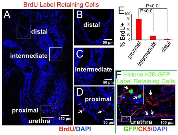
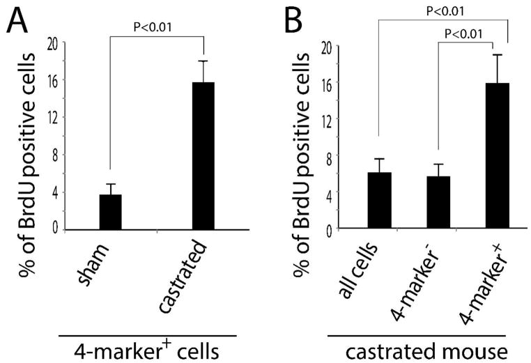
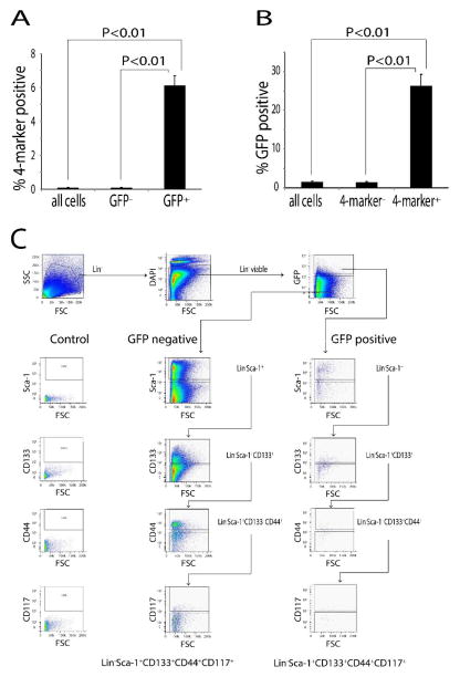
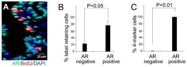
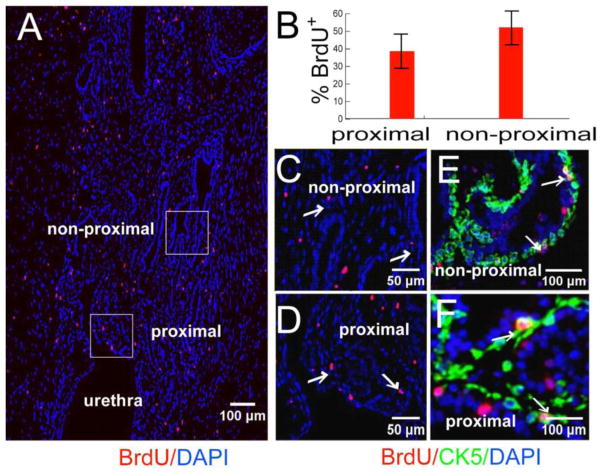
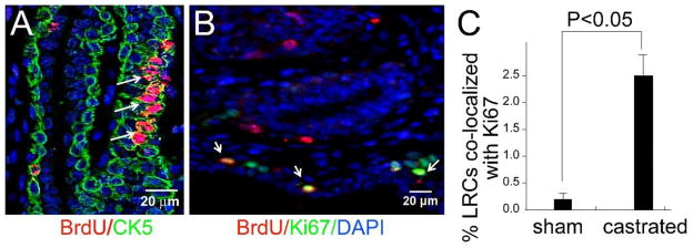
Androgen-deprivation is a mainstay of therapy for advanced prostate cancer but tumor regression is usually incomplete and temporary because of androgen-independent cells in the tumor. It has been speculated that these tumor cells resemble the stem/progenitor cells of the normal prostate. The purpose of this study was to examine the response of slow-cycling progenitor cells in the adult mouse prostate to castration. Proliferating cells in the E16 urogenital sinus were pulse labeled by BrdU administration or by doxycycline-controlled labeling of the histone-H2B GFP mouse. A small population of labeled epithelial cells in the adult prostate localized at the junction of the prostatic ducts and urethra. Fluorescence-activated cell sorting (FACS) showed that GFP label-retaining cells were enriched for cells co-expressing stem cell markers Sca-1, CD133, CD44 and CD117 (4- marker cells; 60-fold enrichment). FACS showed, additionally, that 4-marker cells were androgen receptor positive. Castration induced proliferation and dispersal of E16 labeled cells into more distal ductal segments. When naïve adult mice were administered BrdU daily for 2 weeks after castration, 16% of 4-marker cells exhibited BrdU label in contrast to only 6% of all epithelial cells (P<0.01). In sham-castrated controls less than 4% of 4-marker cells were BrdU labeled (P<0.01). The unexpected and admittedly counter-intuitive finding that castration induced progenitor cell proliferation suggests that androgen deprivation therapy in men with advanced prostate cancer could not only exert pleiotrophic effects on tumor sub-populations but may induce inadvertent expansion of tumor stem cells.
雄激素剥夺疗法是晚期前列腺癌治疗的主要手段,但由于肿瘤中存在雄激素非依赖细胞,肿瘤消退通常不完全且是暂时的。据推测,这些肿瘤细胞类似于正常前列腺的干/祖细胞。本研究的目的是检测成年小鼠前列腺中慢周期祖细胞对去势的反应。通过给予BrdU或利用强力霉素控制标记组蛋白-H2B GFP小鼠,对E16泌尿生殖窦中的增殖细胞进行脉冲标记。成年前列腺中一小部分标记的上皮细胞位于前列腺导管和尿道的交界处。荧光激活细胞分选(FACS)显示,GFP标记保留细胞富含共表达干细胞标志物Sca-1、CD133、CD44和CD117的细胞(四标志物细胞;富集60倍)。此外,FACS显示四标志物细胞为雄激素受体阳性。去势诱导E16标记细胞增殖并扩散到更远端的导管段。当在去势后每天给未处理的成年小鼠注射BrdU,持续2周时,16%的四标志物细胞显示有BrdU标记,而所有上皮细胞中只有6%显示有BrdU标记(P<0.01)。在假去势对照中,少于4%的四标志物细胞有BrdU标记(P<0.01)。去势诱导祖细胞增殖这一意外且违反直觉的发现表明,晚期前列腺癌男性的雄激素剥夺疗法不仅可能对肿瘤亚群产生多效性作用,还可能意外地诱导肿瘤干细胞扩增。