Sun Jing-chuan, Xu Tao, Zuo Qiao, Wang Ruo-bing, Qi Ai-qing, Cao Wen-luo, Sun Ai-jun, Sun Xue-jun, Xu Jiajun
Department of Anatomy, Second Military Medical University, Shanghai, PR China; Graduates Management Unit, Second Military Medical University, Shanghai, PR China.
Department of Ophthalmology, Shanghai Jiaotong University Affiliated Shanghai First People's Hospital, Shanghai, PR China.
PLoS One. 2014 Jun 10;9(6):e99299. doi: 10.1371/journal.pone.0099299. eCollection 2014.
To investigate the effect of molecular hydrogen (H2) in a rat model subjected to optic nerve crush (ONC).
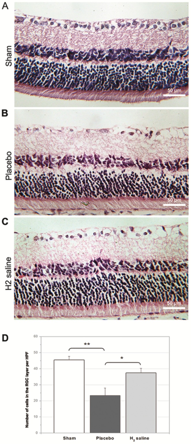
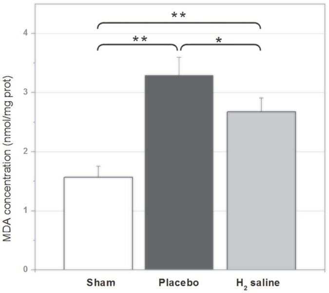
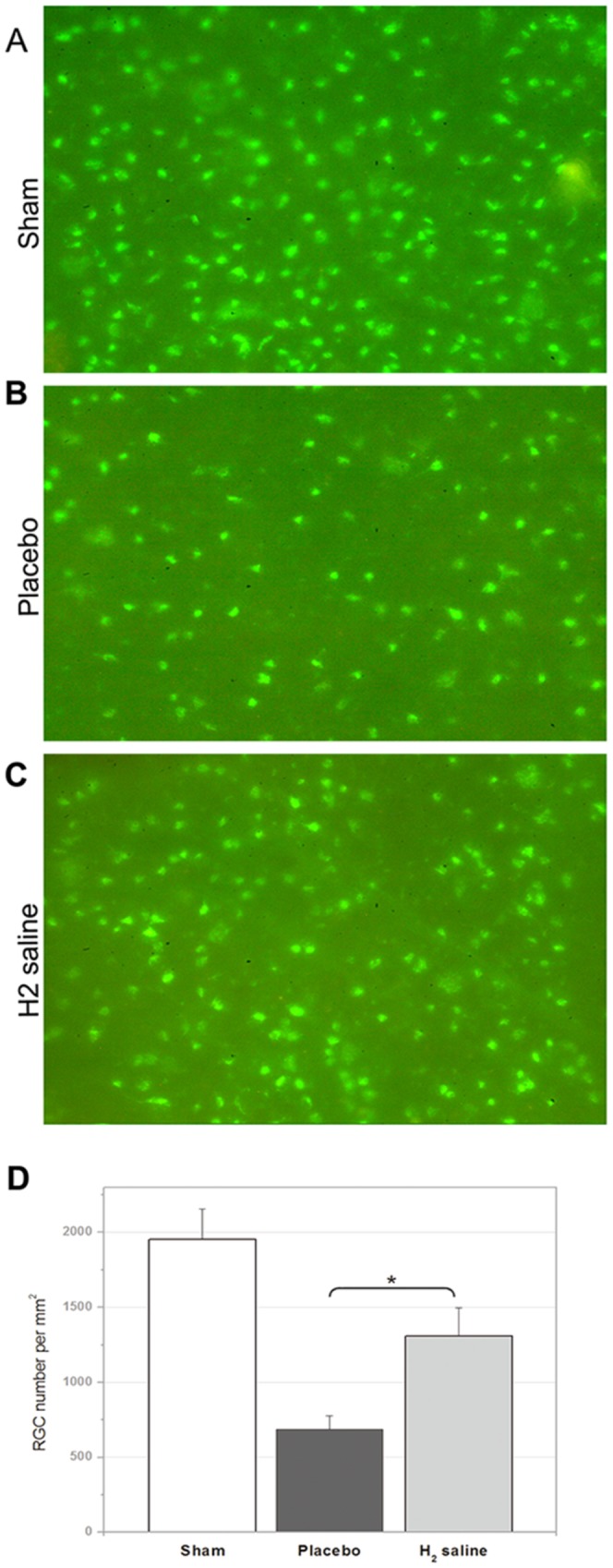
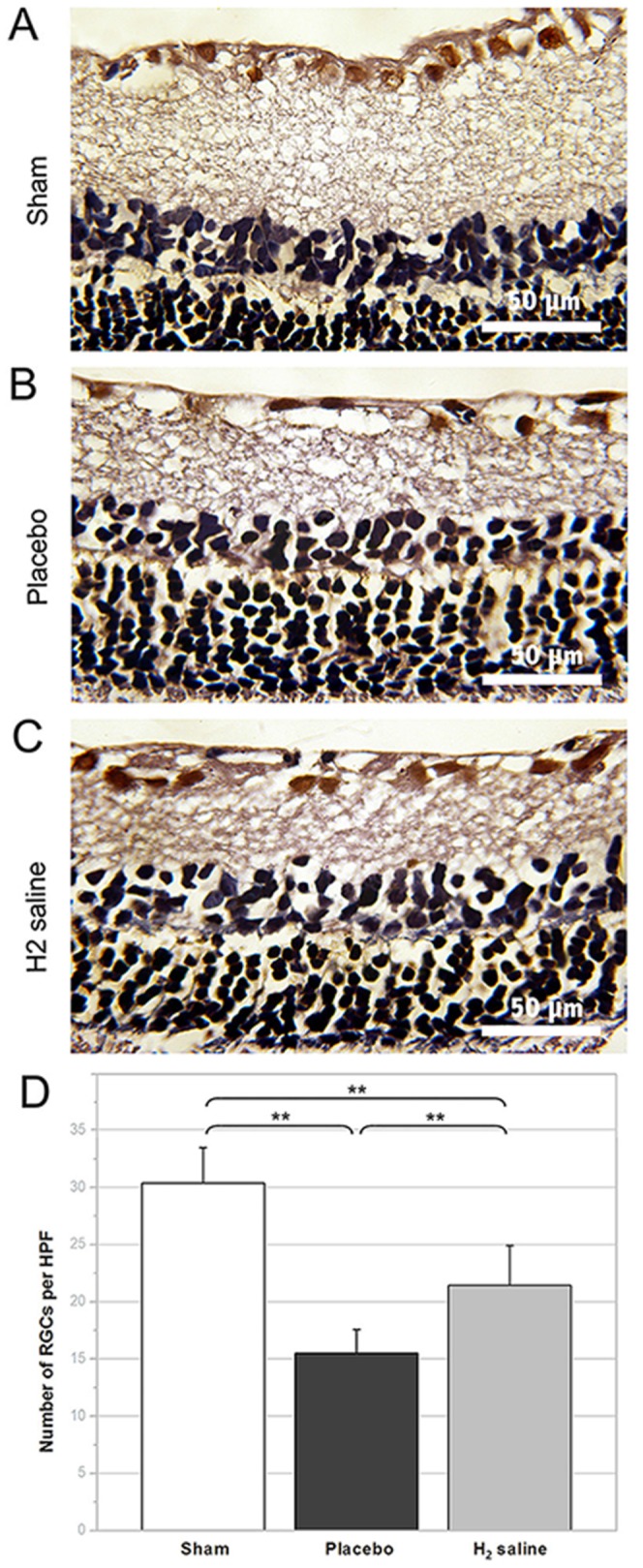
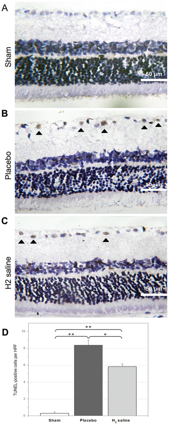
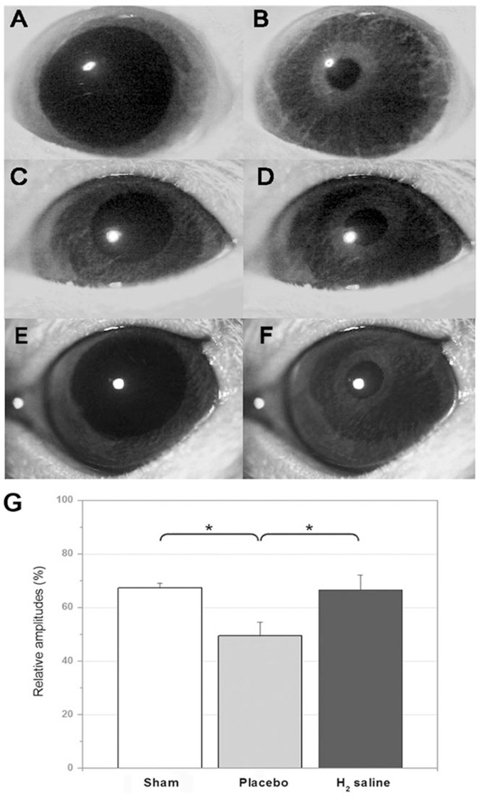
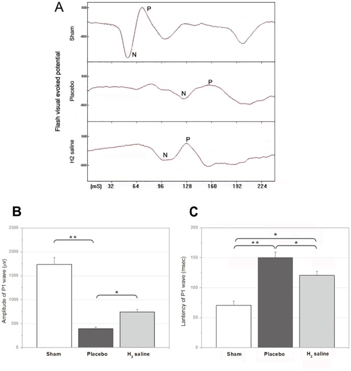
We tested the hypothesis that after optic nerve crush (ONC), retinal ganglion cell (RGC) could be protected by H₂. Rats in different groups received saline or hydrogen-rich saline every day for 14 days after ONC. Retinas from animals in each group underwent measurements of hematoxylin and eosin (H&E) staining, cholera toxin beta (CTB) tracing, gamma synuclein staining, and terminal deoxynucleotidyltransferase-mediated dUTP nick end labeling (TUNEL) staining 2 weeks post operation. Flash visual evoked potentials (FVEP) and pupillary light reflex (PLR) were then tested to evaluate the function of optic nerve. The malondialdehyde (MDA) level in retina was evaluated.
H&E, gamma synuclein staining and CTB tracing showed that the survival rate of RGCs in hydrogen saline-treated group was significantly higher than that in saline-treated group. Apoptosis of RGCs assessed by TUNEL staining were less observed in hydrogen saline-treated group. The MDA level in retina of H₂ group was much lower than that in placebo group. Furthermore, animals treated with hydrogen saline showed better function of optic nerve in assessments of FVEP and PLR.
These results demonstrated that H₂ protects RGCs and helps preserve the visual function after ONC and had a neuroprotective effect in a rat model subjected to ONC.
研究分子氢(H₂)在大鼠视神经挤压伤(ONC)模型中的作用。
我们验证了视神经挤压伤(ONC)后,视网膜神经节细胞(RGC)可被H₂保护这一假设。不同组别的大鼠在ONC后每天接受生理盐水或富氢生理盐水注射,持续14天。术后2周,对每组动物的视网膜进行苏木精-伊红(H&E)染色、霍乱毒素β(CTB)示踪、γ-突触核蛋白染色和末端脱氧核苷酸转移酶介导的dUTP缺口末端标记(TUNEL)染色检测。然后检测闪光视觉诱发电位(FVEP)和瞳孔对光反射(PLR)以评估视神经功能。评估视网膜中的丙二醛(MDA)水平。
H&E染色、γ-突触核蛋白染色和CTB示踪显示,富氢生理盐水治疗组的RGC存活率显著高于生理盐水治疗组。TUNEL染色评估显示,富氢生理盐水治疗组RGC的凋亡较少。H₂组视网膜中的MDA水平远低于安慰剂组。此外,在FVEP和PLR评估中,接受富氢生理盐水治疗的动物视神经功能更好。
这些结果表明,H₂可保护RGC,并有助于在ONC后保留视觉功能,在大鼠ONC模型中具有神经保护作用。