• 文献检索
  • 文档翻译
  • 深度研究
  • 学术资讯
  • Suppr Zotero 插件Zotero 插件
  • 邀请有礼
  • 套餐&价格
  • 历史记录
应用&插件
Suppr Zotero 插件Zotero 插件浏览器插件Mac 客户端Windows 客户端微信小程序
定价
高级版会员购买积分包购买API积分包
服务
文献检索文档翻译深度研究API 文档MCP 服务
关于我们
关于 Suppr公司介绍联系我们用户协议隐私条款
关注我们

Suppr 超能文献

核心技术专利:CN118964589B侵权必究
粤ICP备2023148730 号-1Suppr @ 2026

文献检索

告别复杂PubMed语法,用中文像聊天一样搜索,搜遍4000万医学文献。AI智能推荐,让科研检索更轻松。

立即免费搜索

文件翻译

保留排版,准确专业,支持PDF/Word/PPT等文件格式,支持 12+语言互译。

免费翻译文档

深度研究

AI帮你快速写综述,25分钟生成高质量综述,智能提取关键信息,辅助科研写作。

立即免费体验

用拉曼微光谱术对番茄细胞壁多糖的成像。

Imaging of polysaccharides in the tomato cell wall with Raman microspectroscopy.

机构信息

Institute of Agrophysics, Polish Academy of Sciences, Doswiadczalna 4, 20-290 Lublin, Poland.

出版信息

Plant Methods. 2014 May 29;10:14. doi: 10.1186/1746-4811-10-14. eCollection 2014.

DOI:10.1186/1746-4811-10-14
PMID:24917885
原文链接:https://pmc.ncbi.nlm.nih.gov/articles/PMC4051370/
Abstract

BACKGROUND

The primary cell wall of fruits and vegetables is a structure mainly composed of polysaccharides (pectins, hemicelluloses, cellulose). Polysaccharides are assembled into a network and linked together. It is thought that the percentage of components and of plant cell wall has an important influence on mechanical properties of fruits and vegetables.

RESULTS

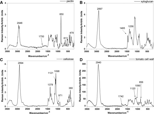
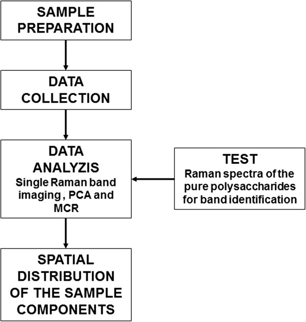
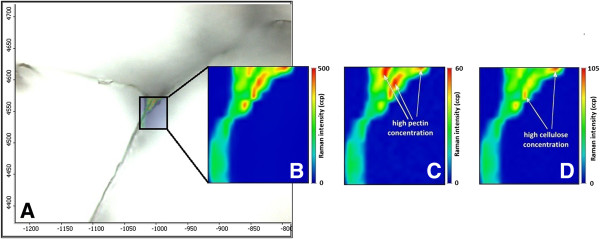
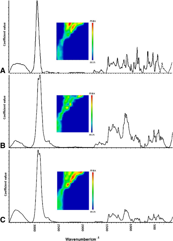
In this study the Raman microspectroscopy technique was introduced to the visualization of the distribution of polysaccharides in cell wall of fruit. The methodology of the sample preparation, the measurement using Raman microscope and multivariate image analysis are discussed. Single band imaging (for preliminary analysis) and multivariate image analysis methods (principal component analysis and multivariate curve resolution) were used for the identification and localization of the components in the primary cell wall.

CONCLUSIONS

Raman microspectroscopy supported by multivariate image analysis methods is useful in distinguishing cellulose and pectins in the cell wall in tomatoes. It presents how the localization of biopolymers was possible with minimally prepared samples.

摘要

背景

水果和蔬菜的初生细胞壁主要由多糖(果胶、半纤维素、纤维素)组成的结构。多糖组装成网络并连接在一起。人们认为成分的百分比和植物细胞壁的百分比对水果和蔬菜的机械性能有重要影响。

结果

本研究将拉曼微光谱技术引入到水果细胞壁中多糖分布的可视化研究中。讨论了样品制备的方法学、拉曼显微镜的测量和多元图像分析。使用单波段成像(初步分析)和多元图像分析方法(主成分分析和多元曲线分辨)来鉴定和定位初生细胞壁中的成分。

结论

多元图像分析支持的拉曼微光谱技术可用于区分番茄细胞壁中的纤维素和果胶。它展示了如何在最小预处理样品的情况下进行生物聚合物的定位。

https://cdn.ncbi.nlm.nih.gov/pmc/blobs/283b/4051370/90f40f499e36/1746-4811-10-14-5.jpg
https://cdn.ncbi.nlm.nih.gov/pmc/blobs/283b/4051370/e017ba557c2b/1746-4811-10-14-1.jpg
https://cdn.ncbi.nlm.nih.gov/pmc/blobs/283b/4051370/ce74f7462c66/1746-4811-10-14-2.jpg
https://cdn.ncbi.nlm.nih.gov/pmc/blobs/283b/4051370/ec713fd0aa9c/1746-4811-10-14-3.jpg
https://cdn.ncbi.nlm.nih.gov/pmc/blobs/283b/4051370/218112195d02/1746-4811-10-14-4.jpg
https://cdn.ncbi.nlm.nih.gov/pmc/blobs/283b/4051370/90f40f499e36/1746-4811-10-14-5.jpg

相似文献

1
Imaging of polysaccharides in the tomato cell wall with Raman microspectroscopy.用拉曼微光谱术对番茄细胞壁多糖的成像。
Plant Methods. 2014 May 29;10:14. doi: 10.1186/1746-4811-10-14. eCollection 2014.
2
Raman imaging of changes in the polysaccharides distribution in the cell wall during apple fruit development and senescence.苹果果实发育和衰老过程中细胞壁多糖分布变化的拉曼成像
Planta. 2016 Apr;243(4):935-45. doi: 10.1007/s00425-015-2456-4. Epub 2016 Jan 5.
3
Hybrid Label-Free Molecular Microscopies for Simultaneous Visualization of Changes in Cell Wall Polysaccharides of Peach at Single- and Multiple-Cell Levels during Postharvest Storage.用于在桃果实采后贮藏过程中在单细胞和多细胞水平上同时可视化细胞壁多糖变化的杂交无标记分子显微镜。
Cells. 2020 Mar 20;9(3):761. doi: 10.3390/cells9030761.
4
Analysis of spatiotemporal changes mechanism of cell wall biopolymers and monosaccharide components in kiwifruit during Botryosphaeria dothidea infection.分析在胶孢炭疽菌侵染猕猴桃过程中细胞壁生物聚合物和单糖组分的时空变化机制。
Spectrochim Acta A Mol Biomol Spectrosc. 2024 Dec 5;322:124837. doi: 10.1016/j.saa.2024.124837. Epub 2024 Jul 22.
5
Use of FT-IR Spectra and PCA to the Bulk Characterization of Cell Wall Residues of Fruits and Vegetables Along a Fraction Process.利用傅里叶变换红外光谱(FT-IR)和主成分分析(PCA)对果蔬细胞壁残渣在分级过程中的整体特性进行表征。
Food Biophys. 2013 Mar;8(1):29-42. doi: 10.1007/s11483-012-9279-7. Epub 2012 Dec 27.
6
Multivariate unmixing approaches on Raman images of plant cell walls: new insights or overinterpretation of results?植物细胞壁拉曼图像的多元解混方法:新见解还是结果的过度解读?
Plant Methods. 2018 Jul 4;14:52. doi: 10.1186/s13007-018-0320-9. eCollection 2018.
7
Properties of cellulose/pectins composites: implication for structural and mechanical properties of cell wall.纤维素/果胶复合材料的性能:对细胞壁结构和力学性能的影响。
Carbohydr Polym. 2012 Oct 1;90(2):1081-91. doi: 10.1016/j.carbpol.2012.06.047. Epub 2012 Jun 21.
8
Pectins, Hemicelluloses and Celluloses Show Specific Dynamics in the Internal and External Surfaces of Grape Berry Skin During Ripening.果胶、半纤维素和纤维素在葡萄浆果成熟过程中,在其果皮的内表面和外表面呈现出特定的动态变化。
Plant Cell Physiol. 2016 Jun;57(6):1332-49. doi: 10.1093/pcp/pcw080. Epub 2016 Apr 19.
9
Combining Raman Imaging and Multivariate Analysis to Visualize Lignin, Cellulose, and Hemicellulose in the Plant Cell Wall.结合拉曼成像与多变量分析以可视化植物细胞壁中的木质素、纤维素和半纤维素。
J Vis Exp. 2017 Jun 10(124):55910. doi: 10.3791/55910.
10
[Studies on Effect of Alkali Pretreatment on Anaerobic Digestion of Rice Straw with Confocal Raman Microscopy].[碱预处理对稻草厌氧消化影响的共聚焦拉曼显微镜研究]
Guang Pu Xue Yu Guang Pu Fen Xi. 2015 Mar;35(3):657-62.

引用本文的文献

1
Integrating Raman spectroscopy and optical meters for nitrogen management in broccoli seedlings.整合拉曼光谱和光学测量仪用于西兰花幼苗的氮素管理
Front Plant Sci. 2025 Jul 18;16:1613503. doi: 10.3389/fpls.2025.1613503. eCollection 2025.
2
Raman and FT-IR Spectroscopy Coupled with Machine Learning for the Discrimination of Different Vegetable Crop Seed Varieties.拉曼光谱和傅里叶变换红外光谱结合机器学习用于鉴别不同蔬菜作物种子品种
Plants (Basel). 2025 Apr 25;14(9):1304. doi: 10.3390/plants14091304.
3
Extracellular Vesicles from Different Mesenchymal Stem Cell Types Exhibit Distinctive Surface Protein Profiling and Molecular Characteristics: A Comparative Analysis.

本文引用的文献

1
Determination of the elastic properties of tomato fruit cells with an atomic force microscope.利用原子力显微镜测定番茄果实细胞的弹性特性。
Sensors (Basel). 2013 Sep 11;13(9):12175-91. doi: 10.3390/s130912175.
2
Use of FT-IR Spectra and PCA to the Bulk Characterization of Cell Wall Residues of Fruits and Vegetables Along a Fraction Process.利用傅里叶变换红外光谱(FT-IR)和主成分分析(PCA)对果蔬细胞壁残渣在分级过程中的整体特性进行表征。
Food Biophys. 2013 Mar;8(1):29-42. doi: 10.1007/s11483-012-9279-7. Epub 2012 Dec 27.
3
Changes in the distribution of cell wall polysaccharides in early fruit pericarp and ovule, from fruit set to early fruit development, in tomato (Solanum lycopersicum).
不同间充质干细胞类型的细胞外囊泡具有独特的表面蛋白谱和分子特征:一项比较分析。
Int J Mol Sci. 2025 Apr 4;26(7):3393. doi: 10.3390/ijms26073393.
4
Shedding new light on the hidden complexity of seeds: chemically selective imaging of seed coats with stimulated Raman scattering microscopy.揭示种子隐藏的复杂性:利用受激拉曼散射显微镜对种皮进行化学选择性成像。
Analyst. 2025 Jan 27;150(3):498-504. doi: 10.1039/d4an01409j.
5
The Correlation Between the Chemical Composition and the Microstructure of the Polysaccharides of Two Varieties of Mexican Red Prickly Pear Fruits.两种墨西哥红刺梨果实多糖的化学成分与微观结构之间的相关性
Foods. 2024 Dec 4;13(23):3914. doi: 10.3390/foods13233914.
6
Imaging Metabolic Flow of Water in Plants with Isotope-Traced Stimulated Raman Scattering Microscopy.利用同位素示踪受激拉曼散射显微镜对植物中水分代谢流进行成像。
Adv Sci (Weinh). 2024 Nov;11(42):e2407543. doi: 10.1002/advs.202407543. Epub 2024 Sep 20.
7
Untapped Potential of Side Stream Products from the Industrial Processing of Fruits: The Biosorption of Anthocyanins on Raspberry Seeds.水果工业加工副产物的未开发潜力:树莓籽对花色苷的生物吸附作用
Foods. 2024 Jul 25;13(15):2334. doi: 10.3390/foods13152334.
8
Expanding the horizons of levan: from microbial biosynthesis to applications and advanced detection methods.扩展低聚糖的应用领域:从微生物生物合成到应用和先进的检测方法。
World J Microbiol Biotechnol. 2024 May 24;40(7):214. doi: 10.1007/s11274-024-04023-w.
9
Tracking deuterium uptake in hydroponically grown maize roots using correlative helium ion microscopy and Raman micro-spectroscopy.使用相关氦离子显微镜和拉曼显微光谱法追踪水培玉米根中氘的吸收情况。
Plant Methods. 2023 Jul 14;19(1):71. doi: 10.1186/s13007-023-01040-y.
10
Vibrational and electrochemical studies of pectin-a candidate towards environmental friendly lithium-ion battery development.果胶用于环保型锂离子电池开发的振动和电化学研究
PNAS Nexus. 2022 Jul 25;1(4):pgac127. doi: 10.1093/pnasnexus/pgac127. eCollection 2022 Sep.
番茄(Solanum lycopersicum)果实从坐果到早期发育过程中,早期果皮和胚珠细胞壁多糖的分布变化。
J Plant Res. 2013 Sep;126(5):719-28. doi: 10.1007/s10265-013-0555-5. Epub 2013 Mar 2.
4
Multivariate curve resolution: a review of advanced and tailored applications and challenges.多元曲线分辨:高级和定制应用及挑战的综述。
Anal Chim Acta. 2013 Feb 26;765:28-36. doi: 10.1016/j.aca.2012.12.028. Epub 2012 Dec 25.
5
The relation of apple texture with cell wall nanostructure studied using an atomic force microscope.利用原子力显微镜研究苹果质地与细胞壁纳米结构的关系。
Carbohydr Polym. 2013 Jan 30;92(1):128-37. doi: 10.1016/j.carbpol.2012.08.103. Epub 2012 Sep 7.
6
Imaging of plant cell walls by confocal Raman microscopy.利用共聚焦拉曼显微镜对植物细胞壁进行成像。
Nat Protoc. 2012 Sep;7(9):1694-708. doi: 10.1038/nprot.2012.092. Epub 2012 Aug 23.
7
Properties of cellulose/pectins composites: implication for structural and mechanical properties of cell wall.纤维素/果胶复合材料的性能:对细胞壁结构和力学性能的影响。
Carbohydr Polym. 2012 Oct 1;90(2):1081-91. doi: 10.1016/j.carbpol.2012.06.047. Epub 2012 Jun 21.
8
A revised architecture of primary cell walls based on biomechanical changes induced by substrate-specific endoglucanases.基于底物特异性内切葡聚糖酶诱导的生物力学变化的初生细胞壁的修订结构。
Plant Physiol. 2012 Apr;158(4):1933-43. doi: 10.1104/pp.111.192880. Epub 2012 Feb 23.
9
Sensing the structural differences in cellulose from apple and bacterial cell wall materials by Raman and FT-IR spectroscopy.通过拉曼和傅里叶变换红外光谱法感知苹果和细菌细胞壁材料中纤维素的结构差异。
Sensors (Basel). 2011;11(6):5543-60. doi: 10.3390/s110605543. Epub 2011 May 25.
10
Functional plant cell wall design revealed by the Raman imaging approach.拉曼成像方法揭示的功能性植物细胞壁设计。
Planta. 2011 Apr;233(4):763-72. doi: 10.1007/s00425-010-1338-z. Epub 2011 Jan 1.