Schmälter Ann-Kristin, Kuzyk Alexandra, Righolt Christiaan H, Neusser Michaela, Steinlein Ortrud K, Müller Stefan, Mai Sabine
Manitoba Institute of Cell Biology, University of Manitoba, Cancer Care Manitoba, 675 McDermot Avenue, Winnipeg, Manitoba, Canada.
BMC Cell Biol. 2014 Jun 12;15:22. doi: 10.1186/1471-2121-15-22.
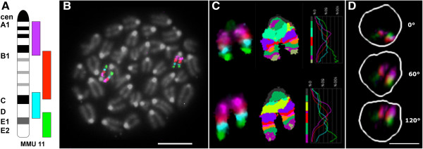
Characterizing the nuclear orientation of chromosomes in the three-dimensional (3D) nucleus by multicolor banding (mBANDing) is a new approach towards understanding nuclear organization of chromosome territories. An mBANDing paint is composed of multiple overlapping subchromosomal probes that represent different regions of a single chromosome. In this study, we used it for the analysis of chromosome orientation in 3D interphase nuclei. We determined whether the nuclear orientation of the two chromosome 11 homologs was random or preferential, and if it was conserved between diploid mouse Pre B lymphocytes of BALB/c origin and primary B lymphocytes of congenic [T38HxBALB/c]N wild-type mice. The chromosome orientation was assessed visually and through a semi-automated quantitative analysis of the radial and angular orientation patterns observed in both B cell types.
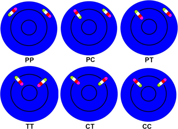
Our data indicate that there are different preferential patterns of chromosome 11 orientation, which are not significantly different between both mouse cell types (p > 0.05). In the most common case for both cell types, both copies of chromosome 11 were oriented in parallel with the nuclear border. The second most common pattern in both types of B lymphocytes was with one homolog of chromosome 11 positioned with its telomeric end towards the nuclear center and with its centromeric end towards the periphery, while the other chromosome 11 was found parallel with the nuclear border. In addition to these two most common orientations present in approximately 50% of nuclei from each cell type, other orientations were observed at lower frequencies.
We conclude that there are probabilistic, non-random orientation patterns for mouse chromosome 11 in the mouse B lymphocytes we investigated (p < 0.0001).
通过多色带型分析(mBANDing)来表征三维(3D)细胞核中染色体的核定位,是一种理解染色体区域核组织的新方法。一种mBANDing探针由多个重叠的亚染色体探针组成,这些探针代表一条单一染色体的不同区域。在本研究中,我们将其用于分析三维间期细胞核中的染色体定位。我们确定了两条11号染色体同源染色体的核定位是随机的还是有偏好的,以及在源自BALB/c的二倍体小鼠前B淋巴细胞和同基因[T38HxBALB/c]N野生型小鼠的原代B淋巴细胞之间这种定位是否保守。通过直观观察以及对两种B细胞类型中观察到的径向和角度定位模式进行半自动定量分析,来评估染色体定位。
我们的数据表明,11号染色体存在不同的偏好定位模式,这两种小鼠细胞类型之间没有显著差异(p > 0.05)。在两种细胞类型最常见的情况下,两条11号染色体均与核边界平行定位。在两种类型的B淋巴细胞中第二常见的模式是,一条11号染色体同源染色体的端粒末端朝向核中心,着丝粒末端朝向核周边,而另一条11号染色体与核边界平行。除了这两种在每种细胞类型约50%的细胞核中出现的最常见定位外,还观察到其他频率较低的定位。
我们得出结论,在我们研究的小鼠B淋巴细胞中,小鼠11号染色体存在概率性的、非随机的定位模式(p < 0.0001)。