Hao Li, Pan Meng-Shu, Zheng Yun, Wang Rui-Feng
Department of Nephrology, The Second Affiliated Hospital of Anhui Medical University, Hefei, Anhui 230601, P.R. China.
Exp Ther Med. 2014 Jun;7(6):1465-1470. doi: 10.3892/etm.2014.1670. Epub 2014 Apr 7.
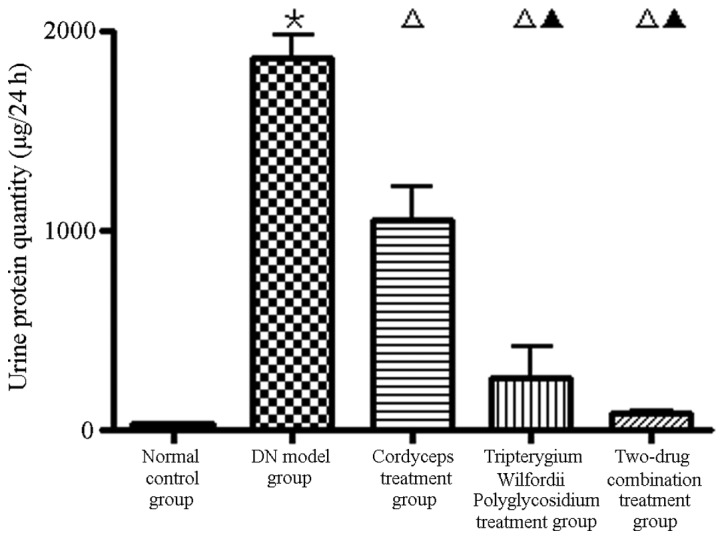
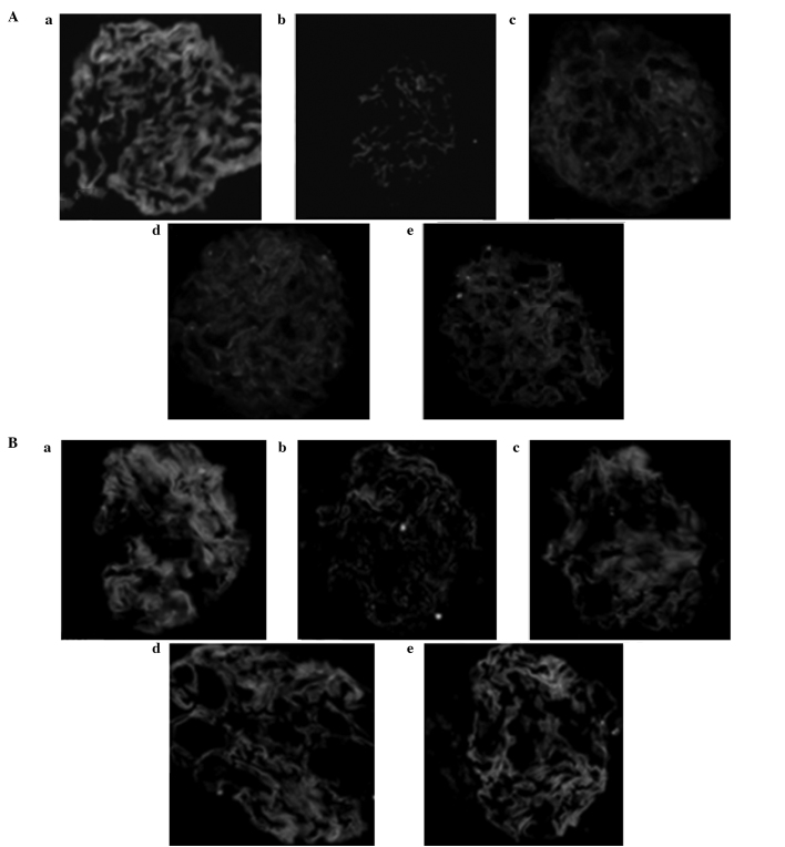
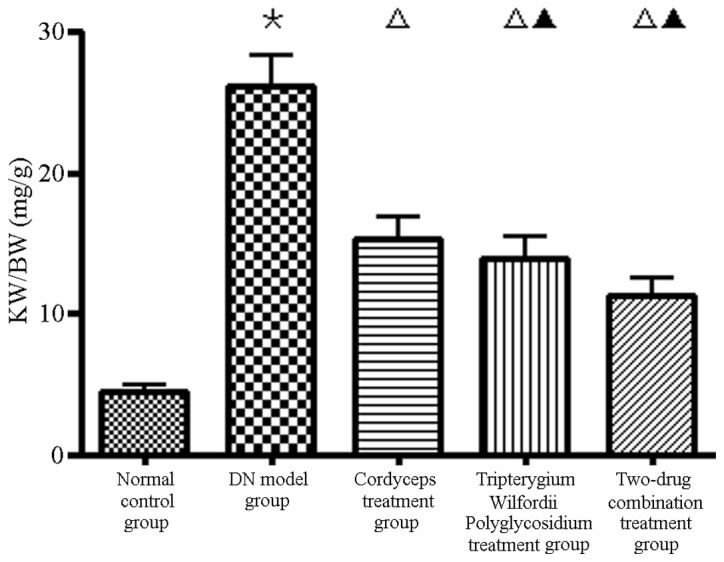
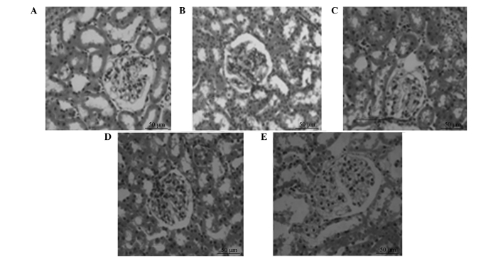
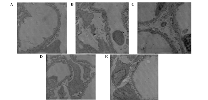
The aim of the present study was to investigate the effects of (CS) and polyglycosidium (TWP) on podocytes in rats with diabetic nephropathy (DN). DN rat models were established and divided randomly into normal control (group A), DN (group B), CS (group C), TWP (group D) and CS and TWP groups (group E). After 12 weeks, levels of 24-h urinary protein, blood urea nitrogen (BUN), serum creatinine (SCR), white blood cells, blood glucose (GLU), aspartate aminotransferase, alanine aminotransferase and kidney weight (KW)/body weight (BW) were determined. Renal pathological changes were evaluated using hematoxylin and eosin staining, whereas the structural changes in the podocytes were observed under a transmission electron microscope. The expression levels of nephrin and podocin were evaluated using immunofluorescence staining. Compared with group A, the SCR and BUN levels in group B were higher (P<0.05) and the GLU, KW/BW and the 24-h urine protein were markedly higher (P<0.01). Moreover, incidences of glomerular disorders, chronic tubulointerstitial damage and glomerular podocyte lesions in groups B, C, D and E were observed, compared with group A. The high cortical expression of nephrin and podocin protein decreased. Compared with group B, the KW/BW and 24-h urinary protein level in groups C, D and E were lower (P<0.01). The glomeruli, tubules and podocytes exhibited pathomorphological improvements and the nephrin and podocin protein expression levels were higher in the nephridial tissue. A decrease in KW/BW and the 24-h urinary protein level, as well as improvements in glomerular disorder, chronic tubulointerstitial damage and glomerular podocyte lesions, were observed in groups C, D and E. Therefore, the results demonstrated that CS and TWP exhibited a protective effect on the podocytes of rats with DN. Moreover, CS combined with TWP increased this protective effect.
本研究旨在探讨虫草菌粉(CS)和糖肾平(TWP)对糖尿病肾病(DN)大鼠足细胞的影响。建立DN大鼠模型并随机分为正常对照组(A组)、DN组(B组)、CS组(C组)、TWP组(D组)以及CS与TWP联合组(E组)。12周后,测定24小时尿蛋白、血尿素氮(BUN)、血清肌酐(SCR)、白细胞、血糖(GLU)、天冬氨酸转氨酶、丙氨酸转氨酶以及肾重(KW)/体重(BW)水平。采用苏木精-伊红染色评估肾脏病理变化,同时在透射电子显微镜下观察足细胞的结构变化。采用免疫荧光染色评估nephrin和podocin的表达水平。与A组相比,B组的SCR和BUN水平更高(P<0.05),GLU、KW/BW以及24小时尿蛋白显著更高(P<0.01)。此外,观察到B、C、D和E组与A组相比出现肾小球病变、慢性肾小管间质损伤和肾小球足细胞病变的发生率。肾皮质中nephrin和podocin蛋白的高表达降低。与B组相比,C、D和E组的KW/BW和24小时尿蛋白水平更低(P<0.01)。肾小球、肾小管和足细胞呈现病理形态学改善,肾组织中nephrin和podocin蛋白表达水平更高。C、D和E组观察到KW/BW和24小时尿蛋白水平降低,以及肾小球病变、慢性肾小管间质损伤和肾小球足细胞病变得到改善。因此,结果表明CS和TWP对DN大鼠的足细胞具有保护作用。此外,CS与TWP联合增强了这种保护作用。