Institute of Genetic Engineering, Southern Medical University, Guangzhou 510515, China.
Department of Urinary Surgery, Nanfang Hosptial, Southern Medical University, Guangzhou 510515, China.
Cancer Cell Int. 2014 Jun 6;14:46. doi: 10.1186/1475-2867-14-46. eCollection 2014.
Pin2/TRF1 binding protein X1 (PinX1) has been identified as an endogenous telomerase inhibitor and a major haploinsufficient tumor suppressor gene. Increasing evidence suggests that reduced expression of PinX1 plays a key role in tumorigenesis. However, the PinX1 expression status and its correlation with the clinicopathological features in prostate cancer (PCa) have not been investigated.
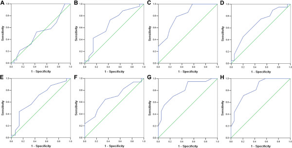
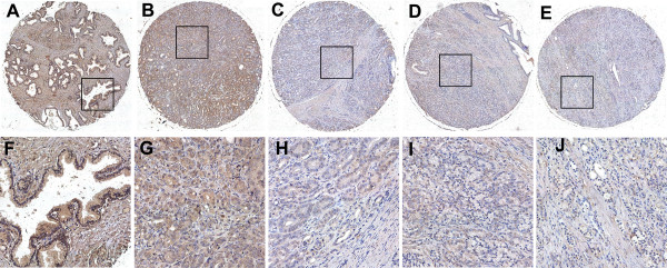
PinX1 mRNA and protein expression in PCa and adjacent normal prostate tissues were evaluated by real-time quantitative RT-PCR (qRT-PCR) and western blotting. The clinicopathological significance of PinX1 was investigated by immunohistochemistry (IHC) analysis on a PCa tissue microarray (TMA). The cut-off score for positive expression of PinX1 was determined by the receiver operating characteristic (ROC) analysis. The correlation between PinX1 expression and clinicopathological features of PCa was analyzed by Chi-square test.
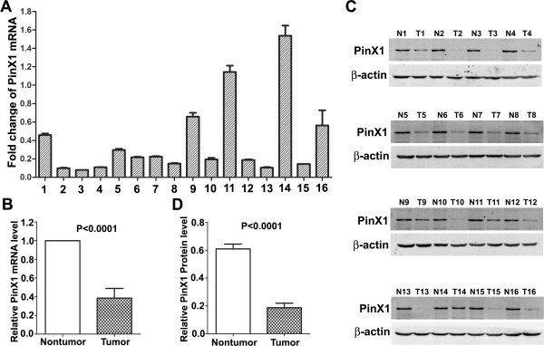
Reduced expression of PinX1 mRNA and protein was observed in the majority of PCa, compared with their paired adjacent normal prostate tissues. When PinX1 positive expression percentage was determined to be above 60% (area under ROC curve = 0.833, P = 0.000), positive expression of PinX1 was observed in 100% (8/8) of normal prostate tissues and 32.5% (13/40) of PCa tissues by IHC. Reduced expression of PinX1 in patients was correlated with advanced clinical stage (χ(2) = 10.230, p = 0.017), high Gleason score (χ(2) = 4.019, p = 0.045), positive regional lymph node metastasis (χ(2) = 10.852, p = 0.004) and distant metastasis (χ(2) = 7.965, p = 0.005).
Our findings suggest that reduced expression of PinX1 is correlates to progressive features in patients with PCa and may serve as a potential marker for diagnosis.
Pin2/TRF1 结合蛋白 X1(PinX1)已被确定为内源性端粒酶抑制剂和主要的杂合不足肿瘤抑制基因。越来越多的证据表明,PinX1 的表达降低在肿瘤发生中起着关键作用。然而,PinX1 的表达状态及其与前列腺癌(PCa)临床病理特征的相关性尚未得到研究。
通过实时定量 RT-PCR(qRT-PCR)和 Western blot 评估 PCa 及相邻正常前列腺组织中的 PinX1mRNA 和蛋白表达。通过前列腺癌组织微阵列(TMA)的免疫组织化学(IHC)分析研究 PinX1 的临床病理意义。通过接收者操作特征(ROC)分析确定 PinX1 阳性表达的截断分数。通过卡方检验分析 PinX1 表达与 PCa 临床病理特征的相关性。
与配对的相邻正常前列腺组织相比,大多数 PCa 中观察到 PinX1mRNA 和蛋白表达降低。当确定 PinX1 阳性表达百分比高于 60%(ROC 曲线下面积=0.833,P=0.000)时,IHC 观察到 100%(8/8)的正常前列腺组织和 32.5%(13/40)的 PCa 组织中 PinX1 阳性表达。患者中 PinX1 的表达降低与临床分期较晚(χ(2)=10.230,p=0.017)、Gleason 评分较高(χ(2)=4.019,p=0.045)、局部淋巴结转移阳性(χ(2)=10.852,p=0.004)和远处转移(χ(2)=7.965,p=0.005)相关。
我们的研究结果表明,PinX1 的表达降低与 PCa 患者的进行性特征相关,可能作为诊断的潜在标志物。