Feng Yu-Zhen, Zhang Qing-Yan, Fu Mei-Ting, Zhang Zhen-Fei, Wei Min, Zhou Jue-Yu, Shi Rong
Institute of Genetic Engineering, School of Basic Medical Sciences, Southern Medical University, Guangzhou, Guangdong 510515, P.R. China.
The First Clinical Medical College, Southern Medical University, Guangzhou, Guangdong 510515, P.R. China.
Oncol Rep. 2017 Jul;38(1):109-119. doi: 10.3892/or.2017.5696. Epub 2017 Jun 2.
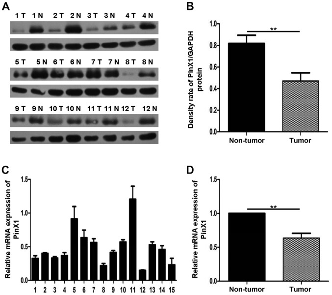
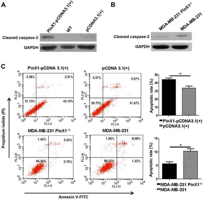
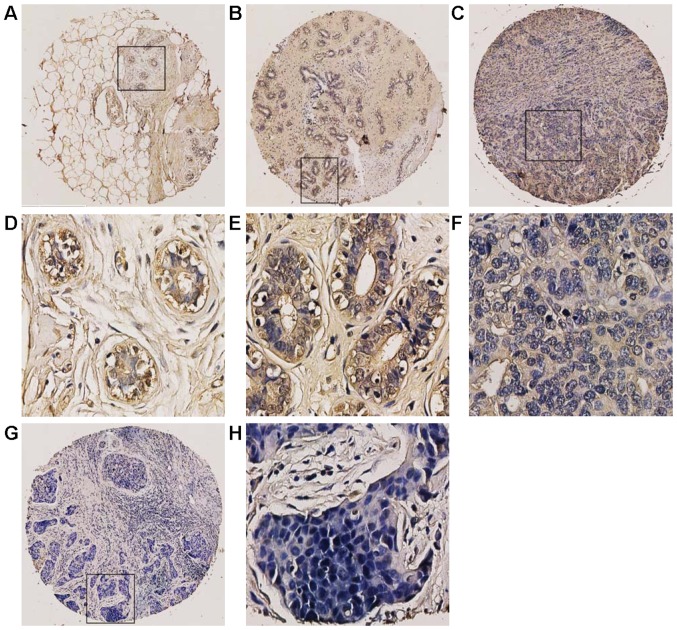
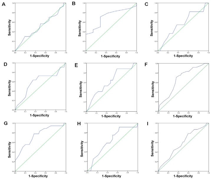
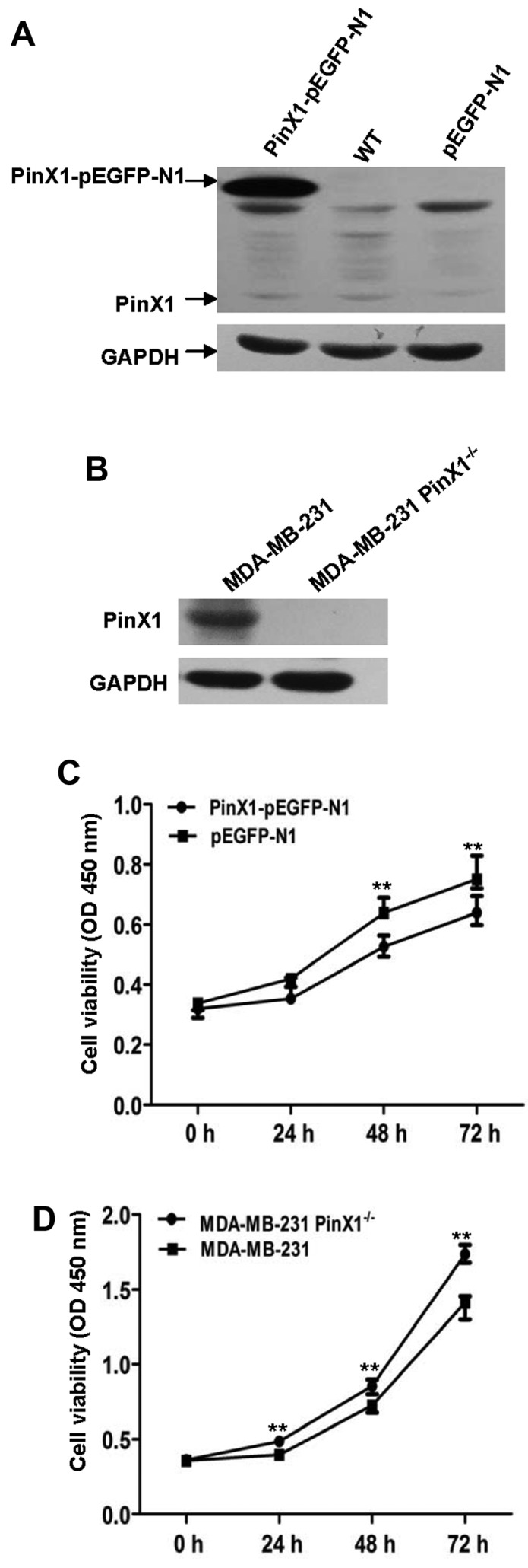
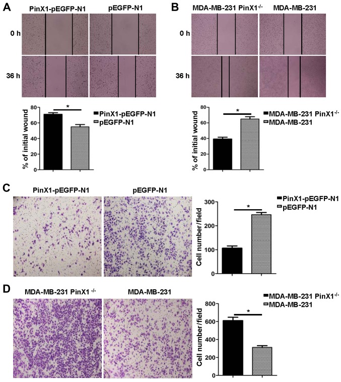
Human Pinx1 protein, associated with shelterin proteins, is widely revealed as a haploinsufficient tumor suppressor. Growing evidence has manifested the deregulation of PinX1 in distinct cancers. Nonetheless, the loss status of PinX1 and its diagnostic, prognostic and clinicopathological significance in Basal-like breast cancer are still unclear. In the present study, the PinX1 expression levels of breast cancer tissues were investigated by qRT-PCR and immunoblotting assays. Then immunohistochemistry (IHC) was performed to detect PinX1 expression on a tissue microarray. The optimal threshold for PinX1 positivity was determined by receiver operating characteristic (ROC) curve analysis. To clarify the probable role of PinX1 in BLBC, the PinX1 knockout and stably over-expressed MDA-MB-231 cell lines were constructed by the CRISPR-Cas9 system and gene transfection. The association of PinX1 expression with cell proliferation, migration and apoptosis of MDA-MB-231 cells were observed by CCK-8 assay, wound healing assay, transwell assay, flow cytometric analysis and immunoblotting of the cleaved caspase-3 protein level. Our results showed that both PinX1 mRNA and protein expression were downregulated in breast cancer tissues (P<0.05). In IHC analysis, the optimal cut-off parameter for PinX1 positive expression was 62.5% (the AUC was 0.749, P<0.01). PinX1 positivity was 76.9% (10/14) in luminal subtypes, 50% (5/10) in Her2-enriched breast cancer and 27.3% (9/33) in basal-like subtypes. Besides, in 59 invasive ductal breast carcinomas, PinX1 expression was inversely related to histology grade (P<0.05) while it was positively associated with PR status (P<0.05) and ER status (P<0.05). These results indicated that low expression of PinX1 correlated with aggressive clinicopathological significance of breast cancer, especially in the basal-like subtype. Besides, we identified that overexpression of PinX1 inhibited the proliferation rates and migration ability and increased the apoptosis rates of BLBC. Our findings demonstrated that low expression of PinX1 was associated with malignant behaviors in basal-like subtype of breast cancer. PinX1 is likely a feasible biomarker and molecular target of BLBC.
与保护帽蛋白相关的人类Pinx1蛋白被广泛揭示为一种单倍剂量不足的肿瘤抑制因子。越来越多的证据表明PinX1在不同癌症中存在失调。然而,PinX1在基底样乳腺癌中的缺失状态及其诊断、预后和临床病理意义仍不清楚。在本研究中,通过qRT-PCR和免疫印迹分析研究了乳腺癌组织中PinX1的表达水平。然后进行免疫组织化学(IHC)检测组织芯片上PinX1的表达。通过受试者操作特征(ROC)曲线分析确定PinX1阳性的最佳阈值。为了阐明PinX1在基底样乳腺癌(BLBC)中可能的作用,利用CRISPR-Cas9系统和基因转染构建了PinX1基因敲除和稳定过表达的MDA-MB-231细胞系。通过CCK-8检测、伤口愈合检测、Transwell检测、流式细胞术分析以及对裂解的caspase-3蛋白水平进行免疫印迹,观察PinX1表达与MDA-MB-231细胞增殖、迁移和凋亡的关系。我们的结果显示,乳腺癌组织中PinX1的mRNA和蛋白表达均下调(P<0.05)。在免疫组织化学分析中,PinX1阳性表达的最佳截断参数为62.5%(曲线下面积为0.749,P<0.01)。在管腔亚型中PinX1阳性率为76.9%(10/14),在人表皮生长因子受体2(Her2)富集型乳腺癌中为50%(5/10),在基底样亚型中为27.3%(9/33)。此外,在59例浸润性导管癌中,PinX1表达与组织学分级呈负相关(P<0.05),而与孕激素受体(PR)状态呈正相关(P<0.05)以及与雌激素受体(ER)状态呈正相关(P<0.05)。这些结果表明,PinX1低表达与乳腺癌侵袭性临床病理特征相关,尤其是在基底样亚型中。此外,我们发现PinX1过表达抑制了基底样乳腺癌的增殖率和迁移能力,并增加了其凋亡率。我们的研究结果表明,PinX1低表达与基底样亚型乳腺癌的恶性行为相关。PinX1可能是基底样乳腺癌一个可行的生物标志物和分子靶点。