• 文献检索
  • 文档翻译
  • 深度研究
  • 学术资讯
  • Suppr Zotero 插件Zotero 插件
  • 邀请有礼
  • 套餐&价格
  • 历史记录
应用&插件
Suppr Zotero 插件Zotero 插件浏览器插件Mac 客户端Windows 客户端微信小程序
定价
高级版会员购买积分包购买API积分包
服务
文献检索文档翻译深度研究API 文档MCP 服务
关于我们
关于 Suppr公司介绍联系我们用户协议隐私条款
关注我们

Suppr 超能文献

核心技术专利:CN118964589B侵权必究
粤ICP备2023148730 号-1Suppr @ 2026

文献检索

告别复杂PubMed语法,用中文像聊天一样搜索,搜遍4000万医学文献。AI智能推荐,让科研检索更轻松。

立即免费搜索

文件翻译

保留排版,准确专业,支持PDF/Word/PPT等文件格式,支持 12+语言互译。

免费翻译文档

深度研究

AI帮你快速写综述,25分钟生成高质量综述,智能提取关键信息,辅助科研写作。

立即免费体验

计算机辅助检测系统在自动化全乳腺超声检查中对肿块的检测:有效性的开发和评估。

Computer-aided detection system for masses in automated whole breast ultrasonography: development and evaluation of the effectiveness.

机构信息

Department of Radiology, Ewha Womans University Mokdong Hospital, Ewha Womans University School of Medicine, Seoul, Korea.

Department of Radiology and Research Institute of Radiology, Asan Medical Center, University of Ulsan College of Medicine, Seoul, Korea.

出版信息

Ultrasonography. 2014 Apr;33(2):105-15. doi: 10.14366/usg.13023. Epub 2014 Feb 26.

DOI:10.14366/usg.13023
PMID:24936503
原文链接:https://pmc.ncbi.nlm.nih.gov/articles/PMC4058980/
Abstract

PURPOSE

The aim of this study was to evaluate the performance of a proposed computer-aided detection (CAD) system in automated breast ultrasonography (ABUS).

METHODS

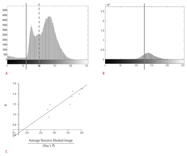
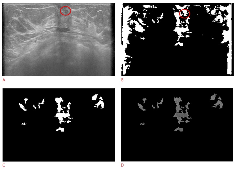
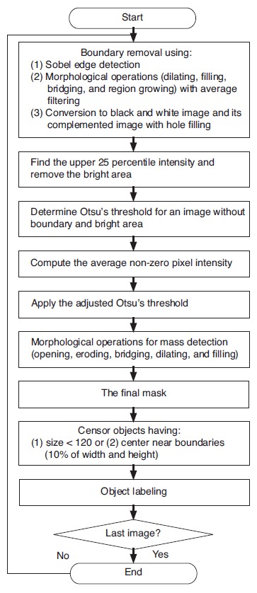
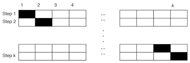
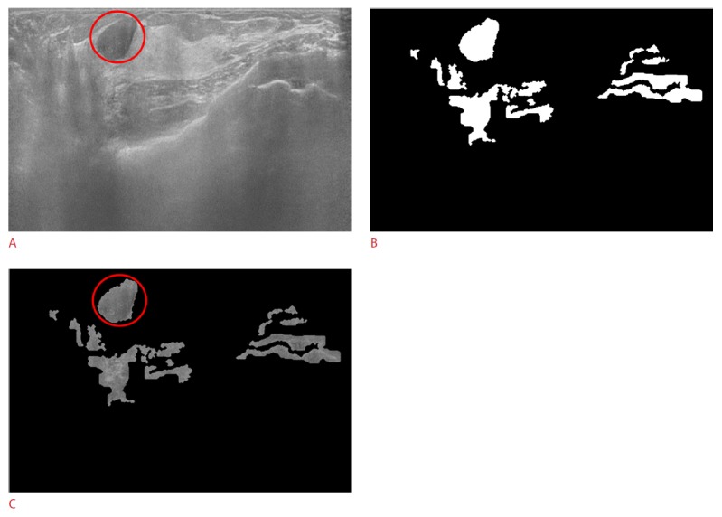
Eighty-nine two-dimensional images (20 cysts, 42 benign lesions, and 27 malignant lesions) were obtained from 47 patients who underwent ABUS (ACUSON S2000). After boundary detection and removal, we detected mass candidates by using the proposed adjusted Otsu's threshold; the threshold was adaptive to the variations of pixel intensities in an image. Then, the detected candidates were segmented. Features of the segmented objects were extracted and used for training/testing in the classification. In our study, a support vector machine classifier was adopted. Eighteen features were used to determine whether the candidates were true lesions or not. A five-fold cross validation was repeated 20 times for the performance evaluation. The sensitivity and the false positive rate per image were calculated, and the classification accuracy was evaluated for each feature.

RESULTS

In the classification step, the sensitivity of the proposed CAD system was 82.67% (SD, 0.02%). The false positive rate was 0.26 per image. In the detection/segmentation step, the sensitivities for benign and malignant mass detection were 90.47% (38/42) and 92.59% (25/27), respectively. In the five-fold cross-validation, the standard deviation of pixel intensities for the mass candidates was the most frequently selected feature, followed by the vertical position of the centroids. In the univariate analysis, each feature had 50% or higher accuracy.

CONCLUSION

The proposed CAD system can be used for lesion detection in ABUS and may be useful in improving the screening efficiency.

摘要

目的

本研究旨在评估一种拟议的计算机辅助检测(CAD)系统在自动乳腺超声(ABUS)中的性能。

方法

从 47 名接受 ABUS(ACUSON S2000)检查的患者中获得 89 个二维图像(20 个囊肿、42 个良性病变和 27 个恶性病变)。边界检测和去除后,我们使用所提出的调整后的 Otsu 阈值来检测肿块候选物;该阈值适应图像中像素强度的变化。然后,检测候选物被分割。分割对象的特征被提取并用于分类的训练/测试。在我们的研究中,采用了支持向量机分类器。采用 18 个特征来确定候选物是否为真正的病变。进行了 20 次五次交叉验证以进行性能评估。计算了每个图像的灵敏度和假阳性率,并评估了每个特征的分类准确性。

结果

在分类步骤中,所提出的 CAD 系统的灵敏度为 82.67%(SD,0.02%)。假阳性率为每幅图像 0.26。在检测/分割步骤中,良性和恶性肿块检测的灵敏度分别为 90.47%(38/42)和 92.59%(25/27)。在五次交叉验证中,肿块候选物的强度像素的标准差是最常被选择的特征,其次是质心的垂直位置。在单变量分析中,每个特征的准确性都在 50%或更高。

结论

所提出的 CAD 系统可用于 ABUS 中的病变检测,可能有助于提高筛查效率。

https://cdn.ncbi.nlm.nih.gov/pmc/blobs/264e/4058980/77d77aa5096f/usg-13023-f6.jpg
https://cdn.ncbi.nlm.nih.gov/pmc/blobs/264e/4058980/04a77c2b8cad/usg-13023-f1.jpg
https://cdn.ncbi.nlm.nih.gov/pmc/blobs/264e/4058980/d12a800d5e3d/usg-13023-f2.jpg
https://cdn.ncbi.nlm.nih.gov/pmc/blobs/264e/4058980/920a5b4fbddc/usg-13023-f3.jpg
https://cdn.ncbi.nlm.nih.gov/pmc/blobs/264e/4058980/68b94e621216/usg-13023-f4.jpg
https://cdn.ncbi.nlm.nih.gov/pmc/blobs/264e/4058980/c85831d6dfec/usg-13023-f5.jpg
https://cdn.ncbi.nlm.nih.gov/pmc/blobs/264e/4058980/77d77aa5096f/usg-13023-f6.jpg

相似文献

1
Computer-aided detection system for masses in automated whole breast ultrasonography: development and evaluation of the effectiveness.计算机辅助检测系统在自动化全乳腺超声检查中对肿块的检测:有效性的开发和评估。
Ultrasonography. 2014 Apr;33(2):105-15. doi: 10.14366/usg.13023. Epub 2014 Feb 26.
2
A 2-Phase Merge Filter Approach to Computer-Aided Detection of Breast Tumors on 3-Dimensional Ultrasound Imaging.一种用于三维超声成像中乳腺肿瘤计算机辅助检测的两阶段合并滤波器方法。
J Ultrasound Med. 2020 Dec;39(12):2439-2455. doi: 10.1002/jum.15365. Epub 2020 Jun 21.
3
Computer-aided tumor detection based on multi-scale blob detection algorithm in automated breast ultrasound images.基于多尺度斑点检测算法的自动乳腺超声图像计算机辅助肿瘤检测。
IEEE Trans Med Imaging. 2013 Jul;32(7):1191-200. doi: 10.1109/TMI.2012.2230403. Epub 2012 Dec 10.
4
Computer-aided diagnosis for the classification of breast masses in automated whole breast ultrasound images.计算机辅助诊断在自动全乳腺超声图像中对乳腺肿块的分类。
Ultrasound Med Biol. 2011 Apr;37(4):539-48. doi: 10.1016/j.ultrasmedbio.2011.01.006.
5
Characterization of masses in digital breast tomosynthesis: comparison of machine learning in projection views and reconstructed slices.数字乳腺断层合成中的肿块特征:在投影视图和重建切片中机器学习的比较。
Med Phys. 2010 Jul;37(7):3576-86. doi: 10.1118/1.3432570.
6
Dedicated computer-aided detection software for automated 3D breast ultrasound; an efficient tool for the radiologist in supplemental screening of women with dense breasts.用于自动化三维乳腺超声的专用计算机辅助检测软件;对于致密型乳腺女性补充筛查的放射科医生来说,是一种有效的工具。
Eur Radiol. 2018 Jul;28(7):2996-3006. doi: 10.1007/s00330-017-5280-3. Epub 2018 Feb 7.
7
A method to test the reproducibility and to improve performance of computer-aided detection schemes for digitized mammograms.一种用于测试数字化乳腺X线摄影计算机辅助检测方案的可重复性并提高其性能的方法。
Med Phys. 2004 Nov;31(11):2964-72. doi: 10.1118/1.1806291.
8
Development of a fully automatic scheme for detection of masses in whole breast ultrasound images.一种用于全乳腺超声图像中肿块检测的全自动方案的开发。
Med Phys. 2007 Nov;34(11):4378-88. doi: 10.1118/1.2795825.
9
Computer-aided diagnosis for classifying benign versus malignant thyroid nodules based on ultrasound images: A comparison with radiologist-based assessments.基于超声图像的甲状腺良恶性结节分类的计算机辅助诊断:与放射科医生评估的比较。
Med Phys. 2016 Jan;43(1):554. doi: 10.1118/1.4939060.
10
Computer-aided lesion diagnosis in automated 3-D breast ultrasound using coronal spiculation.基于冠状面棘突征象的自动三维乳腺超声计算机辅助病变诊断
IEEE Trans Med Imaging. 2012 May;31(5):1034-42. doi: 10.1109/TMI.2012.2184549. Epub 2012 Jan 16.

引用本文的文献

1
Comparison of Automated Breast Ultrasound and Hand-Held Breast Ultrasound in the Screening of Dense Breasts.自动乳腺超声与手持乳腺超声在致密乳腺筛查中的比较。
Rev Bras Ginecol Obstet. 2021 Mar;43(3):190-199. doi: 10.1055/s-0040-1722156. Epub 2021 Apr 15.
2
False-negative results on computer-aided detection software in preoperative automated breast ultrasonography of breast cancer patients.乳腺癌患者术前自动乳腺超声检查中计算机辅助检测软件的假阴性结果。
Ultrasonography. 2021 Jan;40(1):83-92. doi: 10.14366/usg.19076. Epub 2020 Mar 24.
3
Fully automated lesion segmentation and visualization in automated whole breast ultrasound (ABUS) images.

本文引用的文献

1
Computer-aided detection of cancer in automated 3-D breast ultrasound.计算机辅助检测自动三维乳腺超声中的癌症。
IEEE Trans Med Imaging. 2013 Sep;32(9):1698-706. doi: 10.1109/TMI.2013.2263389. Epub 2013 May 16.
2
Computer-aided tumor detection based on multi-scale blob detection algorithm in automated breast ultrasound images.基于多尺度斑点检测算法的自动乳腺超声图像计算机辅助肿瘤检测。
IEEE Trans Med Imaging. 2013 Jul;32(7):1191-200. doi: 10.1109/TMI.2012.2230403. Epub 2012 Dec 10.
3
The Automated Breast Volume Scanner (ABVS): initial experiences in lesion detection compared with conventional handheld B-mode ultrasound: a pilot study of 50 cases.
全乳腺自动超声(ABUS)图像中的全自动病灶分割与可视化
Quant Imaging Med Surg. 2020 Mar;10(3):568-584. doi: 10.21037/qims.2020.01.12.
4
Foundation and methodologies in computer-aided diagnosis systems for breast cancer detection.用于乳腺癌检测的计算机辅助诊断系统的基础与方法
EXCLI J. 2017 Feb 20;16:113-137. doi: 10.17179/excli2016-701. eCollection 2017.
5
Automated Detection Algorithm of Breast Masses in Three-Dimensional Ultrasound Images.三维超声图像中乳腺肿块的自动检测算法
Healthc Inform Res. 2016 Oct;22(4):293-298. doi: 10.4258/hir.2016.22.4.293. Epub 2016 Oct 31.
6
Clinical application of S-Detect to breast masses on ultrasonography: a study evaluating the diagnostic performance and agreement with a dedicated breast radiologist.S-Detect在超声检查乳腺肿块中的临床应用:一项评估其诊断性能及与专业乳腺放射科医生诊断一致性的研究
Ultrasonography. 2017 Jan;36(1):3-9. doi: 10.14366/usg.16012. Epub 2016 Apr 14.
7
Screening Ultrasound in Women with Negative Mammography: Outcome Analysis.乳腺钼靶检查阴性女性的超声筛查:结果分析
Yonsei Med J. 2015 Sep;56(5):1352-8. doi: 10.3349/ymj.2015.56.5.1352.
8
Interobserver agreement on the interpretation of automated whole breast ultrasonography.自动化全乳超声检查的解读的观察者间一致性。
Ultrasonography. 2014 Oct;33(4):252-8. doi: 10.14366/usg.14015. Epub 2014 Apr 21.
自动乳腺容积超声成像系统(ABVS):与传统手持 B 型超声相比在病灶检测方面的初步经验:50 例的初步研究。
Int J Womens Health. 2011;3:337-46. doi: 10.2147/IJWH.S23918. Epub 2011 Oct 11.
4
Breast cancers initially detected by hand-held ultrasound: detection performance of radiologists using automated breast ultrasound data.最初通过手持超声检测出的乳腺癌:放射科医生利用自动乳腺超声数据的检测性能
Acta Radiol. 2011 Feb 1;52(1):8-14. doi: 10.1258/ar.2010.100179.
5
Analysis of eighty-one cases with breast lesions using automated breast volume scanner and comparison with handheld ultrasound.自动乳腺容积扫描与手持超声检查 81 例乳腺病变的分析比较。
Eur J Radiol. 2012 May;81(5):873-8. doi: 10.1016/j.ejrad.2011.02.038. Epub 2011 Mar 21.
6
Radiologists' performance in the detection of benign and malignant masses with 3D automated breast ultrasound (ABUS).放射科医生使用 3D 自动乳腺超声 (ABUS) 检测良性和恶性肿块的表现。
Eur J Radiol. 2011 Apr;78(1):99-103. doi: 10.1016/j.ejrad.2011.01.074. Epub 2011 Feb 16.
7
Supplementary screening sonography in mammographically dense breast: pros and cons.乳腺钼靶致密型中补充性筛查超声:利弊。
Korean J Radiol. 2010 Nov-Dec;11(6):589-93. doi: 10.3348/kjr.2010.11.6.589. Epub 2010 Oct 29.
8
Breast cancer detection: radiologists' performance using mammography with and without automated whole-breast ultrasound.乳腺癌检测:使用配有和不配有自动全乳房超声的乳腺 X 线摄影术时放射科医生的表现。
Eur Radiol. 2010 Nov;20(11):2557-64. doi: 10.1007/s00330-010-1844-1. Epub 2010 Jul 15.
9
Rapid image stitching and computer-aided detection for multipass automated breast ultrasound.多通道自动化乳腺超声的快速图像拼接和计算机辅助检测。
Med Phys. 2010 May;37(5):2063-73. doi: 10.1118/1.3377775.
10
Breast cancer detection using automated whole breast ultrasound and mammography in radiographically dense breasts.乳腺 X 线摄影致密型乳腺中应用自动全乳腺超声和乳腺 X 线摄影筛查乳腺癌。
Eur Radiol. 2010 Mar;20(3):734-42. doi: 10.1007/s00330-009-1588-y. Epub 2009 Sep 2.