• 文献检索
  • 文档翻译
  • 深度研究
  • 学术资讯
  • Suppr Zotero 插件Zotero 插件
  • 邀请有礼
  • 套餐&价格
  • 历史记录
应用&插件
Suppr Zotero 插件Zotero 插件浏览器插件Mac 客户端Windows 客户端微信小程序
定价
高级版会员购买积分包购买API积分包
服务
文献检索文档翻译深度研究API 文档MCP 服务
关于我们
关于 Suppr公司介绍联系我们用户协议隐私条款
关注我们

Suppr 超能文献

核心技术专利:CN118964589B侵权必究
粤ICP备2023148730 号-1Suppr @ 2026

文献检索

告别复杂PubMed语法,用中文像聊天一样搜索,搜遍4000万医学文献。AI智能推荐,让科研检索更轻松。

立即免费搜索

文件翻译

保留排版,准确专业,支持PDF/Word/PPT等文件格式,支持 12+语言互译。

免费翻译文档

深度研究

AI帮你快速写综述,25分钟生成高质量综述,智能提取关键信息,辅助科研写作。

立即免费体验

利用 X 射线显微断层扫描技术对染色和天然椎间盘中的 3D 微观结构进行可视化。

Visualising the 3D microstructure of stained and native intervertebral discs using X-ray microtomography.

机构信息

Centre for Doctoral Training in Regenerative Medicine, University of Manchester, Manchester, UK.

Division of Cell Matrix Biology and Regenerative Medicine, University of Manchester, Manchester, UK.

出版信息

Sci Rep. 2017 Nov 24;7(1):16279. doi: 10.1038/s41598-017-16354-w.

DOI:10.1038/s41598-017-16354-w
PMID:29176563
原文链接:https://pmc.ncbi.nlm.nih.gov/articles/PMC5701246/
Abstract

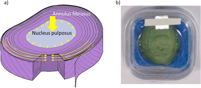
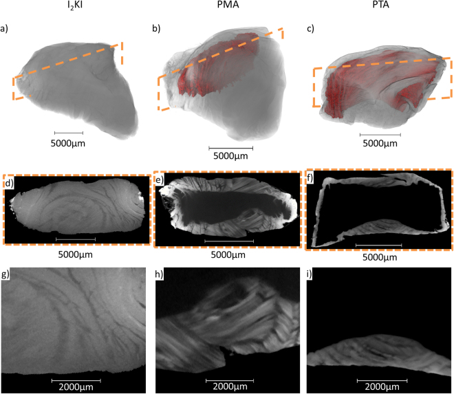
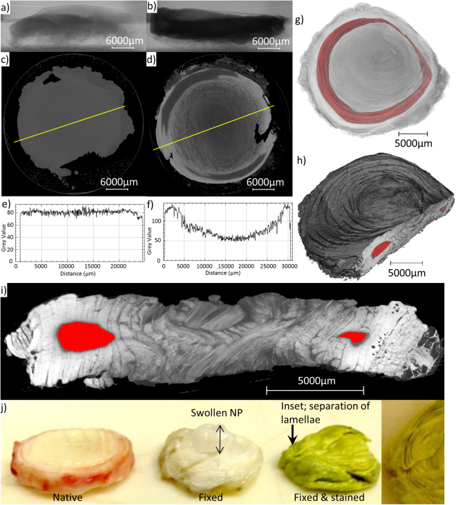
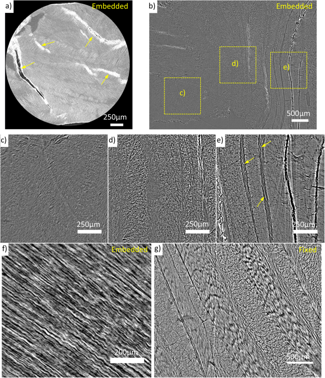
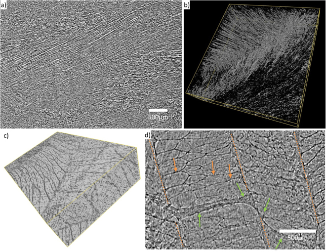
Intervertebral disc degeneration (IVDD) is linked to low back pain. Microstructural changes during degeneration have previously been imaged using 2D sectioning techniques and 3D methods which are limited to small specimens and prone to inducing artefacts from sample preparation. This study explores micro computed X-ray tomography (microCT) methods with the aim of resolving IVD 3D microstructure whilst minimising sample preparation artefacts. Low X-ray absorption contrast in non-mineralised tissue can be enhanced using staining and phase contrast techniques. A step-wise approach, including comparing three stains, was used to develop microCT for bovine tail IVD using laboratory and synchrotron sources. Staining successfully contrasted collagenous structures; however not all regions were stained and the procedure induced macroscopic structural changes. Phase contrast microCT of chemically fixed yet unstained samples resolved the nucleus pulposus, annulus fibrosus and constituent lamellae, and finer structures including collagen bundles and cross-bridges. Using the same imaging methods native tissue scans were of slightly lower contrast but free from sample processing artefacts. In the future these methods may be used to characterise structural remodelling in soft (non-calcified) tissues and to conduct in situ studies of native loaded tissues and constructs to characterise their 3D mechanical properties.

摘要

椎间盘退变(IVDD)与下腰痛有关。以前使用 2D 切片技术和 3D 方法对退变过程中的微观结构变化进行了成像,但这些方法仅限于小样本,并且容易因样本制备而产生伪影。本研究探索了微计算机 X 射线断层扫描(microCT)方法,旨在最小化样本制备伪影的同时解析 IVDD 的 3D 微观结构。非矿化组织中低 X 射线吸收率可以通过染色和相衬技术来增强。本研究采用逐步方法,包括比较三种染色剂,使用实验室和同步辐射源开发了用于牛尾椎间盘的 microCT。染色成功地对比了胶原结构;然而,并非所有区域都被染色,并且该过程会引起宏观结构变化。对化学固定但未染色的样本进行相衬 microCT 可解析核髓、纤维环和组成的薄片以及更精细的结构,包括胶原束和交叉桥。使用相同的成像方法,天然组织扫描的对比度稍低,但没有样本处理伪影。将来,这些方法可用于表征软(未钙化)组织中的结构重塑,并对天然负载组织和构建体进行原位研究,以表征其 3D 机械性能。

https://cdn.ncbi.nlm.nih.gov/pmc/blobs/973d/5701246/ee17f0951659/41598_2017_16354_Fig7_HTML.jpg
https://cdn.ncbi.nlm.nih.gov/pmc/blobs/973d/5701246/b6a602720c28/41598_2017_16354_Fig1_HTML.jpg
https://cdn.ncbi.nlm.nih.gov/pmc/blobs/973d/5701246/ed7b4a9022af/41598_2017_16354_Fig2_HTML.jpg
https://cdn.ncbi.nlm.nih.gov/pmc/blobs/973d/5701246/c6683a7bcd9a/41598_2017_16354_Fig3_HTML.jpg
https://cdn.ncbi.nlm.nih.gov/pmc/blobs/973d/5701246/816c5f450798/41598_2017_16354_Fig4_HTML.jpg
https://cdn.ncbi.nlm.nih.gov/pmc/blobs/973d/5701246/638ffcad4582/41598_2017_16354_Fig5_HTML.jpg
https://cdn.ncbi.nlm.nih.gov/pmc/blobs/973d/5701246/c82147d73550/41598_2017_16354_Fig6_HTML.jpg
https://cdn.ncbi.nlm.nih.gov/pmc/blobs/973d/5701246/ee17f0951659/41598_2017_16354_Fig7_HTML.jpg

相似文献

1
Visualising the 3D microstructure of stained and native intervertebral discs using X-ray microtomography.利用 X 射线显微断层扫描技术对染色和天然椎间盘中的 3D 微观结构进行可视化。
Sci Rep. 2017 Nov 24;7(1):16279. doi: 10.1038/s41598-017-16354-w.
2
A review of techniques for visualising soft tissue microstructure deformation and quantifying strain Ex Vivo.软组织微观结构变形可视化技术及体外应变定量评估研究综述。
J Microsc. 2018 Dec;272(3):165-179. doi: 10.1111/jmi.12701. Epub 2018 Apr 14.
3
Synchrotron tomography of intervertebral disc deformation quantified by digital volume correlation reveals microstructural influence on strain patterns.基于数字体图像相关技术的椎间盘变形同步辐射断层成像定量分析显示了微观结构对变形模式的影响。
Acta Biomater. 2019 Jul 1;92:290-304. doi: 10.1016/j.actbio.2019.05.021. Epub 2019 May 11.
4
The Quantitative Structural and Compositional Analyses of Degenerating Intervertebral Discs Using Magnetic Resonance Imaging and Contrast-Enhanced Micro-Computed Tomography.使用磁共振成像和增强对比微计算机断层扫描对退变椎间盘进行定量结构和成分分析。
Ann Biomed Eng. 2017 Nov;45(11):2626-2634. doi: 10.1007/s10439-017-1891-8. Epub 2017 Jul 25.
5
Three-dimensional characterization of in vivo intervertebral disc degeneration using EPIC-μCT.使用EPIC-μCT对体内椎间盘退变进行三维表征。
Osteoarthritis Cartilage. 2014 Nov;22(11):1918-25. doi: 10.1016/j.joca.2014.08.006. Epub 2014 Aug 20.
6
A novel technique for the contrast-enhanced microCT imaging of murine intervertebral discs.一种用于小鼠椎间盘对比增强微型计算机断层扫描成像的新技术。
J Mech Behav Biomed Mater. 2016 Oct;63:66-74. doi: 10.1016/j.jmbbm.2016.06.003. Epub 2016 Jun 8.
7
Regional variations in discrete collagen fibre mechanics within intact intervertebral disc resolved using synchrotron computed tomography and digital volume correlation.利用同步辐射计算机断层扫描和数字体素相关技术解析完整椎间盘内离散胶原纤维力学的区域变化。
Acta Biomater. 2022 Jan 15;138:361-374. doi: 10.1016/j.actbio.2021.10.012. Epub 2021 Oct 10.
8
Three-dimensional microstructural reconstruction of the ovine intervertebral disc using ultrahigh field MRI.利用超高场 MRI 对羊椎间盘进行三维微观结构重建。
Spine J. 2018 Nov;18(11):2119-2127. doi: 10.1016/j.spinee.2018.06.356. Epub 2018 Jun 30.
9
Comparison of Synchrotron Radiation-based Propagation Phase Contrast Imaging and Conventional Micro-computed Tomography for Assessing Intervertebral Discs and Endplates in a Murine Model.基于同步辐射的传播相位对比成像与传统微计算机断层扫描在评估小鼠模型椎间盘和终板中的比较
Spine (Phila Pa 1976). 2017 Aug 1;42(15):E883-E889. doi: 10.1097/BRS.0000000000002110.
10
Direct microCT imaging of non-mineralized connective tissues at high resolution.直接高分辨率 microCT 成像非矿化结缔组织。
Connect Tissue Res. 2014 Jan-Feb;55(1):52-60. doi: 10.3109/03008207.2013.867333.

引用本文的文献

1
Spatial and temporal evaluation of iodine uptake and radiodensity in meniscus tissue using contrast-enhanced micro-CT.使用对比增强微型计算机断层扫描对半月板组织中碘摄取和放射密度进行空间和时间评估。
Heliyon. 2024 Dec 7;10(24):e41080. doi: 10.1016/j.heliyon.2024.e41080. eCollection 2024 Dec 30.
2
Quantification of porcine lower thoracic spinal cord morphology with intact dura mater using high-resolution μCT.使用高分辨率 μCT 对完整硬脑膜的猪下胸段脊髓形态进行定量分析。
J Neuroimaging. 2024 Nov-Dec;34(6):646-663. doi: 10.1111/jon.13239. Epub 2024 Oct 10.
3
Seeing the unseen: The role of bioimaging techniques for the diagnostic interventions in intervertebral disc degeneration.

本文引用的文献

1
High-contrast X-ray micro-radiography and micro-CT of ex-vivo soft tissue murine organs utilizing ethanol fixation and large area photon-counting detector.利用乙醇固定和大面积光子计数探测器对离体小鼠软组织器官进行高对比度X射线显微放射成像和显微CT成像。
Sci Rep. 2016 Jul 27;6:30385. doi: 10.1038/srep30385.
2
Optimal Contrast Agent Staining of Ligaments and Tendons for X-Ray Computed Tomography.用于X射线计算机断层扫描的韧带和肌腱的最佳造影剂染色
PLoS One. 2016 Apr 14;11(4):e0153552. doi: 10.1371/journal.pone.0153552. eCollection 2016.
3
Optical Coherence Tomographic Elastography Reveals Mesoscale Shear Strain Inhomogeneities in the Annulus Fibrosus.
洞察不可见:生物成像技术在椎间盘退变诊断干预中的作用。
Bone Rep. 2024 Jun 27;22:101784. doi: 10.1016/j.bonr.2024.101784. eCollection 2024 Sep.
4
Visualization of rat tendon in three dimensions using micro-Computed Tomography.使用微型计算机断层扫描对大鼠肌腱进行三维可视化。
MethodsX. 2024 Jan 10;12:102565. doi: 10.1016/j.mex.2024.102565. eCollection 2024 Jun.
5
Does Vitamin K2 Influence the Interplay between Diabetes Mellitus and Intervertebral Disc Degeneration in a Rat Model?维生素 K2 是否影响糖尿病与椎间盘退变在大鼠模型中的相互作用?
Nutrients. 2023 Jun 25;15(13):2872. doi: 10.3390/nu15132872.
6
Heterotopic mineral deposits in intact rat Achilles tendons are characterized by a unique fiber-like structure.完整大鼠跟腱中的异位矿物质沉积具有独特的纤维状结构。
J Struct Biol X. 2023 Feb 24;7:100087. doi: 10.1016/j.yjsbx.2023.100087. eCollection 2023.
7
Importance of Matrix Cues on Intervertebral Disc Development, Degeneration, and Regeneration.基质线索对椎间盘发育、退变和再生的重要性。
Int J Mol Sci. 2022 Jun 21;23(13):6915. doi: 10.3390/ijms23136915.
8
Regional variations in discrete collagen fibre mechanics within intact intervertebral disc resolved using synchrotron computed tomography and digital volume correlation.利用同步辐射计算机断层扫描和数字体素相关技术解析完整椎间盘内离散胶原纤维力学的区域变化。
Acta Biomater. 2022 Jan 15;138:361-374. doi: 10.1016/j.actbio.2021.10.012. Epub 2021 Oct 10.
9
A quality optimization approach to image Achilles tendon microstructure by phase-contrast enhanced synchrotron micro-tomography.基于相衬增强同步辐射微层析成像的图像跟腱微结构的质量优化方法。
Sci Rep. 2021 Aug 27;11(1):17313. doi: 10.1038/s41598-021-96589-w.
10
Engineering multifunctional metal/protein hybrid nanomaterials as tools for therapeutic intervention and high-sensitivity detection.设计多功能金属/蛋白质杂化纳米材料作为治疗干预和高灵敏度检测的工具。
Chem Sci. 2020 Dec 14;12(7):2480-2487. doi: 10.1039/d0sc05215a.
光学相干断层扫描弹性成像揭示了纤维环中的中尺度剪切应变不均匀性。
Spine (Phila Pa 1976). 2016 Jul 1;41(13):E770-E777. doi: 10.1097/BRS.0000000000001463.
4
Assessing the three-dimensional collagen network in soft tissues using contrast agents and high resolution micro-CT: Application to porcine iliac veins.使用造影剂和高分辨率微型计算机断层扫描评估软组织中的三维胶原网络:在猪髂静脉中的应用
C R Biol. 2015 Jul;338(7):425-33. doi: 10.1016/j.crvi.2015.04.009. Epub 2015 May 29.
5
Determining collagen distribution in articular cartilage using contrast-enhanced micro-computed tomography.使用对比增强型微型计算机断层扫描确定关节软骨中的胶原蛋白分布。
Osteoarthritis Cartilage. 2015 Sep;23(9):1613-21. doi: 10.1016/j.joca.2015.05.004. Epub 2015 May 21.
6
Morphological Characterisation of Unstained and Intact Tissue Micro-architecture by X-ray Computed Micro- and Nano-Tomography.通过X射线计算机微纳断层扫描对未染色完整组织微结构进行形态学表征
Sci Rep. 2015 May 15;5:10074. doi: 10.1038/srep10074.
7
A high-throughput system for high-quality tomographic reconstruction of large datasets at Diamond Light Source.一种用于在钻石光源处对大型数据集进行高质量断层扫描重建的高通量系统。
Philos Trans A Math Phys Eng Sci. 2015 Jun 13;373(2043). doi: 10.1098/rsta.2014.0398.
8
Data Analysis WorkbeNch (DAWN).数据分析工作台(DAWN)。
J Synchrotron Radiat. 2015 May;22(3):853-8. doi: 10.1107/S1600577515002283. Epub 2015 Apr 2.
9
ISSLS Prize Winner: A Detailed Examination of the Elastic Network Leads to a New Understanding of Annulus Fibrosus Organization.国际腰椎研究学会奖获得者:对弹性网络的详细研究引发了对纤维环组织结构的新认识。
Spine (Phila Pa 1976). 2015 Aug 1;40(15):1149-57. doi: 10.1097/BRS.0000000000000943.
10
Three dimensional mesoscale analysis of translamellar cross-bridge morphologies in the annulus fibrosus using optical coherence tomography.使用光学相干断层扫描对纤维环中跨层横桥形态进行三维中尺度分析。
J Orthop Res. 2015 Mar;33(3):304-11. doi: 10.1002/jor.22778. Epub 2015 Jan 6.